


Unraveling the Stress-Eczema Link
For years, individuals with eczema have anecdotally reported that psychological stress exacerbates their condition. However, the precise biological mechanism
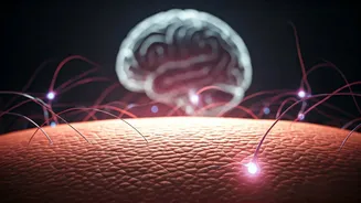
behind this observation has remained elusive until now. Recent scientific investigations have pinpointed a novel brain-to-skin communication pathway. This pathway appears to actively intensify inflammatory responses within the skin by directing nerve signals specifically towards immune cells. This finding moves beyond simply acknowledging stress as a trigger and suggests it is an integral component of the eczema disease process itself. The implications are significant: if this identified pathway can be therapeutically targeted, it could offer complementary approaches to existing eczema treatments, potentially by managing stress more effectively or by developing medications that interrupt these critical nerve signals.
The Neuroimmune Axis Explained
Atopic dermatitis, the most prevalent form of eczema, is a complex condition that extends beyond a mere skin eruption. It is characterized by a compromised skin barrier, persistent inflammation, and an intense, often unbearable itch. This itch can initiate a vicious cycle of scratching, leading to further skin damage and a worsening of symptoms. It is well-established that stress can aggravate this cycle by negatively impacting immune system function, weakening the skin's protective barrier, and heightening the sensation of itch. The skin's unique composition, teeming with both nerve endings and immune cells, makes it a prime location for intricate communication between the nervous and immune systems. Despite this, the precise sequence of events that translates psychological stress into inflammatory flare-ups in eczema has been a persistent question. A key player under scrutiny has been the eosinophil, a type of immune cell strongly implicated in allergic inflammation and severe dermatitis. Eosinophils possess the capacity to release harmful granule proteins and inflammatory substances that can significantly worsen skin damage. What remained unclear was precisely how nerve signals, influenced by stress, manage to draw these eosinophils into the skin and activate their inflammatory functions.
Investigating Stress Pathways
To delve deeper into this complex interaction, researchers led by Jiahe Tian conducted a comprehensive study. They analyzed clinical data from 51 individuals diagnosed with atopic dermatitis and simultaneously employed mouse models to observe the effects of stress on skin inflammation. Their findings revealed a compelling correlation: higher reported stress levels in patients were associated with a greater accumulation of eosinophils within their skin. Furthermore, the study identified a specific group of sympathetic neurons, known as prodynorphin-positive (Pdyn+) noradrenergic neurons, which directly innervate hairy skin. Through further experiments, the team demonstrated that these particular neurons act as conduits, transmitting stress-related signals from the brain down to the skin. Once in the skin, these signals amplify inflammation, and this amplification is critically dependent on the presence and activity of eosinophils. The neurons orchestrate this by recruiting eosinophils via the CCL11-CCR3 signaling pathway and subsequently activating them through the adrenergic receptor beta2 (Adrb2). The research confirmed that inhibiting either these specific neurons or eosinophils effectively reduced stress-induced inflammation. Conversely, stimulating these neurons led to an escalation of inflammation. These discoveries suggest that peripheral sympathetic nerves, rather than the more commonly studied hypothalamus-pituitary-adrenal (HPA) axis, play a pivotal role in mediating this stress response in eczema. Consequently, the stress-induced increase in eosinophils and the direct interface between Pdyn+ sympathetic neurons and eosinophils are emerging as promising targets for future therapeutic interventions in atopic dermatitis.