


Ants: Tiny Titans
Ants, often overlooked, are a cornerstone of our planet's ecosystems, with an estimated 20 quadrillion individuals teeming across the globe. Their societal
structures are complex, and their roles in nature are vital, ranging from environmental engineers to specialized predators. Scientists have long been fascinated by their intricate lives and diverse forms. This fascination has now led to a revolutionary method for studying them, transforming our understanding of these ubiquitous insects. By capturing their internal and external structures with unparalleled clarity, researchers are unlocking new insights into their biology and evolution, proving that even the smallest creatures hold immense scientific value and biological complexity.
Revolutionary Scanning Technique
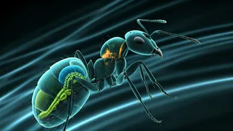
A collaborative effort has produced a stunning collection of high-resolution 3D images of ants, made possible by an intense week of X-ray scanning using a synchrotron particle accelerator. This cutting-edge technology, typically used for fundamental physics research, allowed for the rapid imaging of nearly 2,200 specimens sourced from global collections. Traditional methods would have taken years to achieve similar results. The process involved researchers working around the clock, meticulously positioning and scanning each ant. This accelerated approach enabled the visualization of internal organs like brains, guts, and glands, as well as external features, in remarkable detail. The ability to capture such comprehensive data so quickly marks a significant advancement in entomological study.
From Contorted to Natural Poses
Initial 3D renderings captured the ants in their preserved, often contorted, postures, which did not accurately represent their natural forms. To overcome this, collaborators employed artificial intelligence to automate the process of repositioning the scanned ant models into more lifelike poses. This AI-driven technique has profound implications, allowing for a more intuitive and accurate understanding of ant anatomy and morphology. The potential applications extend far beyond entomology; this method could eventually be adapted to reconstruct the natural poses of any organism, providing invaluable tools for scientists, educators, artists, and animators alike. The combination of advanced imaging and intelligent software is revolutionizing how we study and visualize the natural world.
Global Specimens, Local Scan
The project began with a remarkable feat of specimen collection, with thousands of ant samples meticulously gathered from museums and private collections worldwide. Julian Katzke, a former graduate student, recalled the logistical challenge and surprise of fitting over 2,000 vials, each containing a precious specimen, into just two tote bags for transport to Germany. This highlights a significant advantage of studying small organisms: their sheer volume does not necessitate vast logistical undertakings for data collection. Once at the synchrotron facility, a robotic system efficiently handled the placement of each sample for scanning. Watching these diverse insects materialize on screen in real-time was a profound experience for the researchers, revealing an astonishing array of peculiar forms, specialized mandibles, and unique body structures.
Inside the Tiny Body
Beyond external morphology, the synchrotron scans provided an unprecedented view into the internal anatomy of the ants. Researchers were able to clearly visualize their delicate brains, intricate digestive systems, and various glands. This internal detail is crucial for understanding their physiology and behavior. For instance, the study was able to rapidly survey numerous species and confirm that a mineralized exoskeleton, akin to mollusk shells, previously identified in fungus-farming ants, is a common trait within that group. This comparative internal analysis, done non-destructively, significantly accelerates research and allows for broader discoveries about evolutionary adaptations and biological functions within ant species, opening new avenues for anatomical and physiological research.
Transformative Research Tool
The impact of this new scanning methodology is recognized as transformative by experts in the field. Jessica Ware, a curator at the American Museum of Natural History, notes that researchers can now examine valuable study specimens internally without the need for destructive dissection. This preservation of specimens is a critical benefit for ongoing research. Despite the significant cost associated with synchrotron facilities, Ware is confident that this approach will become more widely adopted due to its immense value. The ability to create detailed digital models of organisms also fosters wider engagement, allowing the public to appreciate the complex and diverse shapes found in nature, making scientific discoveries more accessible and inspiring.