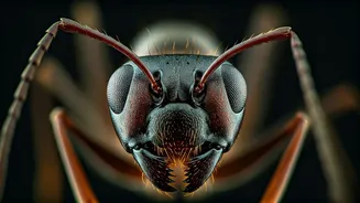


Unveiling Ant Diversity
Ants, a ubiquitous presence in nearly every ecosystem, exhibit an astounding diversity in form and function. From the formidable bullet ant, notorious
for its intensely painful sting, to minuscule predators specializing in consuming spider eggs, their morphological variations are vast. Scientists like Evan Economo emphasize their complex social structures, which, combined with their sheer abundance—estimated at over 20 quadrillion individuals—underscore their significant ecological roles. This fascination with ant diversity has now been amplified by a remarkable new study that has produced breathtakingly detailed 3D images of nearly 2,200 ant specimens. These scans offer an unprecedented glimpse into the intricate anatomy of these creatures, revealing details both externally and internally with exceptional clarity, all made freely available through an online portal called antscan.info.
Accelerated Imaging Breakthrough
The creation of these high-resolution 3D ant models was made possible by a concentrated, week-long scanning effort using a synchrotron particle accelerator located in Germany. This advanced technology allowed researchers, including Thomas van de Kamp from the Karlsruhe Institute of Technology, to process an immense number of specimens—close to 2,200—in a remarkably short period. Traditional methods for achieving such detailed scans would have typically spanned several years. The synchrotron's power lies in its ability to expose samples to intense light, enabling much faster scanning than smaller micro-CT scanners. Crucially, it allows for the visualization of soft tissues without the need for chemical staining agents, a significant advantage for preserving sample integrity and detail.
AI for Natural Poses
A common challenge with preserved specimens is their often contorted or unnatural postures, described by Economo as being "crinkled up." To address this, the research team employed artificial intelligence. Their computer science collaborators developed an AI system capable of automating the repositioning of the scanned ants into more natural, lifelike poses. This innovative technique not only enhances the aesthetic appeal of the ant models but also provides a more accurate representation for scientific study. Economo envisions this AI-driven repositioning being applicable to a broader range of organisms in the future, benefiting scientists, educators, and even artists and animators by creating more realistic digital representations.
Global Collections Assembled
The ambitious project began with a collaboration between Evan Economo and Thomas van de Kamp, stemming from Economo's time at the Okinawa Institute of Science and Technology. Specimens were meticulously gathered from museum collections and private holdings worldwide. Julian Katzke, a former graduate student in Economo's lab, played a key role in transporting over 2,000 vials of ants to Germany, highlighting the remarkable compactness of these small insects—fitting within a manageable suitcase. Inside the synchrotron facility, a robotic arm systematically positioned each sample for scanning, a process observed with great interest by van de Kamp from the control room. The sheer variety observed, from ants with specialized mandibles and spines to those covered in unique hairs, underscored the astonishing differences among species.
Inside the Tiny Anatomy
Beyond their external morphology, the high-resolution scans provided by the synchrotron offered an unprecedented opportunity to examine the internal anatomy of ants. Researchers could now visualize vital organs such as brains, guts, and various glands within these minuscule creatures. This internal view proved invaluable for scientific discovery. For instance, the team could rapidly survey many species and confirm observations about the exoskeletons of fungus-farming ants. Previously, it was known that these ants possessed a mineralized armor resembling mollusk shells; the new scans facilitated a swift confirmation of this trait across numerous related species, demonstrating the power of this imaging technique for comparative studies and uncovering widespread biological features.
Transformative Scientific Potential
Experts outside the study, like Jessica Ware, Curator and Chair of Invertebrate Zoology at the American Museum of Natural History, recognize the transformative potential of this new imaging technology. Ware highlights that researchers can now investigate valuable study organisms internally without the destructive process of dissection. Despite the significant costs associated with synchrotron access, she is confident that this approach will become increasingly adopted within the scientific community due to its unparalleled detail and non-invasive nature. Economo shares this optimism, expressing excitement about extending the application of these techniques beyond ants to encompass a wider array of life forms, potentially enabling the creation of detailed digital worlds and advanced robotics.