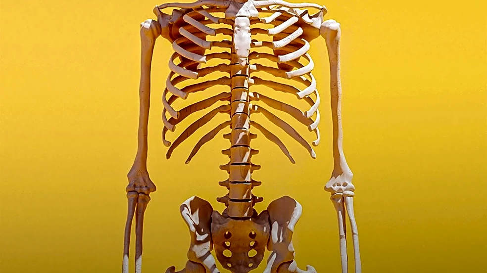








The thoracic vertebrae represent a crucial segment of the human vertebral column, positioned between the cervical vertebrae of the neck and the lumbar vertebrae of the lower back. These twelve vertebrae, often abbreviated T1 through T12, are integral components of both the vertebral column and the bones of the thorax. Their unique structural characteristics are specifically adapted to support the rib cage and protect the spinal cord, while also allowing
for a degree of flexibility and movement essential for daily activities.
Understanding the specific features of the thoracic vertebrae is key to appreciating their role in spinal mechanics and overall body structure. While the first and ninth through twelfth thoracic vertebrae possess certain peculiarities, the general characteristics observed from the second through eighth vertebrae provide a foundational understanding of this spinal region.
General Characteristics of Thoracic Vertebrae
The vertebral bodies in the middle of the thoracic region are distinctly heart-shaped. They are as broad in the anteroposterior direction as they are in the transverse direction, contributing to their robust nature. These bodies are slightly thicker behind than in front, and they feature flat upper and lower surfaces. From side to side, they are convex in front and deeply concave behind, with a slight constriction laterally and anteriorly. A notable feature on either side of these vertebral bodies are two costal demi-facets. One demi-facet is located superiorly, near the root of the pedicle, while the other is inferiorly positioned, in front of the inferior vertebral notch. In a fresh state, these demi-facets are covered with cartilage and, when the vertebrae articulate, they form oval surfaces with the intervertebral fibrocartilages, designed to receive the heads of the ribs.
The pedicles of the thoracic vertebrae are directed backward and slightly upward. The inferior vertebral notches are notably large and deeper than those found in any other region of the vertebral column, a characteristic that further distinguishes this segment. The laminae, which are broad and thick, are imbricated, meaning they overlap those of the subjacent vertebrae much like tiles on a roof. This overlapping arrangement, along with their connection to the pedicles, serves to surround and protect the delicate spinal cord within.
Specialized Features and Ligamentous Support
The intervertebral foramen in the thoracic region is small and circular. There are two such foramina at each intervertebral level, one for the right and one for the left exiting nerve roots, allowing for the passage of spinal nerves. Posterior to the vertebral body is the vertebral foramen, also known as the spinal canal, which is a large opening that contains and protects the spinal cord at the thoracic level.
The spinous process of a thoracic vertebra is long and triangular in coronal section. It projects obliquely downward from the lamina and terminates in a tuberculated extremity. These processes exhibit a significant overlap from the fifth to the eighth vertebrae, though they are less oblique in direction both above and below this central range. The superior articular processes are thin plates of bone that project upward from the junctions of the pedicles and laminae. Their articular facets are practically flat and are directed backward, slightly lateralward, and upward. Conversely, the inferior articular processes are largely fused with the laminae and extend slightly beyond their lower borders, with their facets directed proximally, medially, and inferiorly to the lamina.
Further contributing to the stability and controlled movement of the thoracic spine are the transverse processes. These processes originate from the arch behind the superior articular processes and pedicles. They are thick, strong, and of considerable length, directed obliquely backward and lateralward. Each transverse process ends in a clubbed extremity, on the front of which is a small, concave surface designed for articulation with the tubercle of a rib. The intertransverse ligaments, which are weak and sheet-like, interconnect adjacent transverse processes in the thoracic spine. These ligaments play a crucial role in limiting lateral flexion and rotation of the spine, thereby contributing to the overall stability and controlled mobility of this vital segment of the vertebral column.