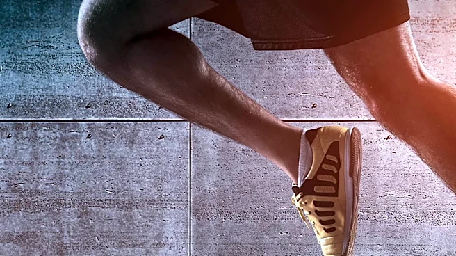








The rectus femoris muscle is a crucial component of the human leg, specifically one of the four quadriceps muscles. Situated prominently in the middle of the front of the thigh, this muscle plays a vital role in both hip and knee movements. Its unique structure and neurological connections allow for complex actions like extending the leg at the knee joint and flexing the thigh at the hip joint, making it indispensable for daily activities and athletic
endeavors. Understanding its anatomy and function provides insight into the intricate mechanics of the human musculoskeletal system.
Anatomical Structure and Connections
The rectus femoris muscle is characterized by its fusiform shape, meaning it is spindle-like, wider in the middle and tapering at both ends. Its superficial fibers are arranged in a bipenniform manner, resembling a feather with muscle fibers attaching obliquely to a central tendon, while its deep fibers run straight down to a deep aponeurosis. This muscle originates from two distinct points on the hip bone: an anterior or straight tendon arises from the anterior inferior iliac spine, and a posterior or reflected tendon comes from a groove above the rim of the acetabulum. These two tendons converge at an acute angle and expand into an aponeurosis that extends downward on the anterior surface of the muscle, from which the muscular fibers emerge.
The muscle terminates in a broad and thick aponeurosis that covers the lower two-thirds of its posterior surface. This aponeurosis gradually narrows into a flattened tendon, which then inserts into the base of the patella, or kneecap. This insertion point is shared with the other three quadriceps muscles—the vastus medialis, vastus intermedius, and vastus lateralis—all of which attach to the patella via the quadriceps tendon. The rectus femoris receives its blood supply from the descending branch of the lateral femoral circumflex artery, ensuring it has the necessary nutrients and oxygen to perform its functions.
Neurological Control and Function
The voluntary contraction of the rectus femoris muscle is a complex process initiated in the brain. The neurons responsible for voluntary thigh contraction originate near the summit of the medial side of the precentral gyrus, which is the primary motor area of the brain. These upper motor neurons send a nerve signal that travels down the corticospinal tract, passing through the brainstem and spinal cord. The signal moves through the internal capsule, the cerebral peduncle, and into the medulla. In the medullary pyramid, the corticospinal tract crosses over to the opposite side of the body, becoming the lateral corticospinal tract.
The nerve signal continues its journey down the lateral corticospinal tract until it reaches spinal nerve L4. At this point, the signal synapses from the upper motor neurons to the lower motor neurons. The signal then travels through the anterior root of L4 and into the anterior rami of the L4 nerve, exiting the spinal cord through the lumbar plexus. The posterior division of the L4 root forms the femoral nerve, which is responsible for innervating the quadriceps femoris, including the rectus femoris. When the rectus femoris receives this signal, it contracts, resulting in the extension of the knee and the flexion of the thigh at the hip. This intricate neural pathway highlights the precise control the brain exerts over muscle movement.
Functional Dynamics and Antagonism
The rectus femoris is a powerful muscle, but its effectiveness in certain actions can be influenced by its length. For instance, it acts as a weaker hip flexor when the knee is extended. This is due to a phenomenon known as active insufficiency, where the muscle is already shortened, limiting its ability to generate maximum force. In such cases, other hip flexors like the iliacus, psoas major, and tensor fasciae latae contribute more significantly to the action. Similarly, the rectus femoris is not the primary muscle for knee extension when the hip is already flexed, again due to active insufficiency.
Conversely, the muscle's ability to flex the hip and extend the knee can be compromised in a position of full hip extension and knee flexion. This is known as passive insufficiency, where the muscle is stretched to its maximum length, preventing further movement. The rectus femoris also has a direct antagonistic relationship with the hamstrings, both at the hip and the knee joints. This means that when the rectus femoris contracts to perform an action, the hamstrings typically relax, and vice versa, allowing for coordinated and controlled movement. This interplay between agonist and antagonist muscles is fundamental to the biomechanics of the leg.