



The gastrocnemius muscle, while prominent as part of the calf, is not an isolated entity. It exists within a complex network of muscles, tendons, and nerves in the lower leg, forming crucial relationships that enable its diverse functions. Understanding these connections, including its shared tendon with the soleus and its innervation by the tibial nerve, provides a more complete picture of its role in human locomotion and its susceptibility to certain
conditions.
The Triceps Surae: A Unified Calf Muscle
The gastrocnemius is located superficially in the posterior compartment of the leg, meaning it is closer to the skin. Beneath it, or farther from the skin, lies the soleus muscle. Together, the gastrocnemius and the soleus form a significant portion of what is commonly known as the calf muscle. In fact, some anatomists consider these two muscles to be a single functional unit, referring to them as the triceps surae, or the "three-headed muscle of the calf." This perspective arises because they share a common insertion point via the Achilles tendon, also known as the calcaneal tendon, which attaches to the heel bone.
This shared tendon is a critical anatomical feature, allowing both muscles to contribute to plantar flexing the foot at the ankle joint. The close relationship extends further with the plantaris muscle and a portion of its tendon running between the gastrocnemius and soleus. The plantaris muscle is involved in "locking" the knee from a standing position, highlighting the interconnectedness of these lower leg structures.
Neurological Pathways and Clinical Vulnerabilities
The gastrocnemius muscle's function is entirely dependent on its innervation, which comes from the tibial nerve. This nerve is a branch of the sciatic nerve, which itself originates from the sacral plexus, specifically from the anterior rami of Lumbar 4-5 and Sacral 1-5. The signal for muscle contraction begins in the precentral gyrus of the brain's cerebrum, travels down through upper motor neurons, crosses in the medulla oblongata, and then synapses with lower motor neurons in the spinal cord before reaching the tibial nerve and, subsequently, the gastrocnemius.
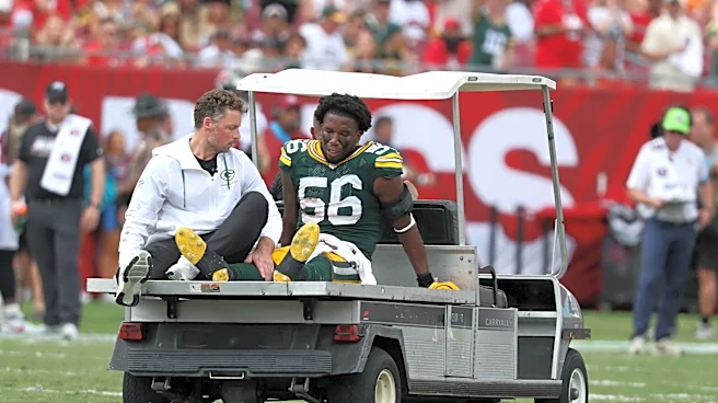
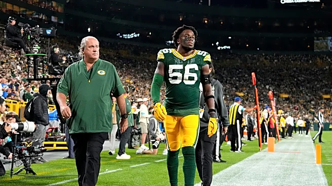
Given its active role in movement, the gastrocnemius is susceptible to various clinical issues. It is prone to spasms, which are painful, involuntary contractions that can last for several minutes. A severe ankle dorsiflexion force can lead to a Medial Gastrocnemius Strain (MGS), commonly referred to as a "torn" or "strained" calf muscle, causing acute pain and disability. Overuse can also lead to inflammation of the muscle. Furthermore, anatomical abnormalities involving the medial head of the gastrocnemius muscle can result in popliteal artery entrapment syndrome, a condition that can affect blood flow to the lower leg. These clinical considerations underscore the importance of understanding the muscle's anatomy and its surrounding structures.