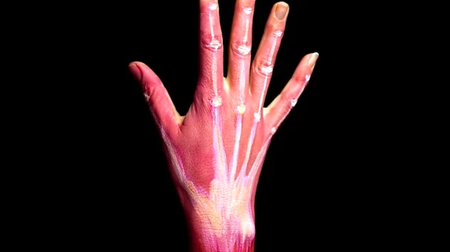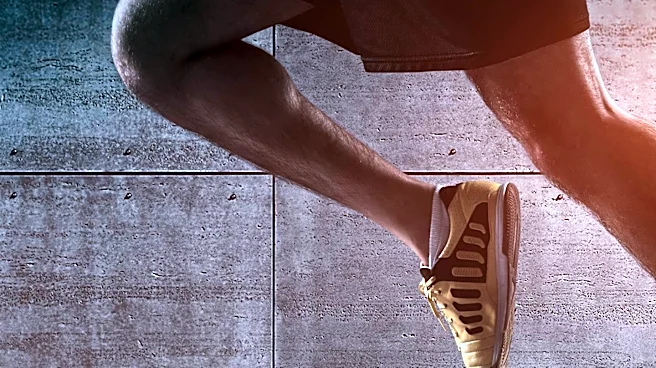





The vastus lateralis, also known as the vastus externus, stands as the largest and most powerful component of the quadriceps femoris muscle group, located in the thigh. This muscle plays a crucial role in the mechanics of the human leg, primarily responsible for extending the knee joint. Its function allows for the forward movement of the lower leg, a fundamental action in walking, running, and many other daily activities. Beyond its primary role in movement,
the vastus lateralis holds significant importance in the medical field, particularly as a recommended site for intramuscular injections in specific patient populations.
Anatomical Origins and Connections
The vastus lateralis muscle has a complex and extensive origin, arising from multiple areas of the femur, the large bone in the thigh. Its origins include the upper part of the intertrochanteric line, which is a ridge located between the greater and lesser trochanters of the femur. It also originates from the lower, anterior borders of the greater trochanter, a large, irregular eminence at the upper part of the femur, and extends to the outer border of the gluteal tuberosity. Furthermore, the upper half of the outer border of the linea aspera, a rough line on the posterior surface of the femur, contributes to its origin.
These various points of origin converge to form an aponeurosis, which is a broad, flat tendon. This aponeurosis effectively covers the upper three-quarters of the vastus lateralis muscle. From the inner surface of this aponeurosis, numerous muscle fibers emerge, contributing to the bulk of the muscle. Additional fibers also arise from the tendon of the gluteus maximus muscle and from the septum that separates the vastus lateralis from the short head of the biceps femoris, further illustrating its intricate anatomical connections within the thigh.
Insertion and Role in the Quadriceps Tendon
The fibers of the vastus lateralis muscle coalesce into a substantial fleshy mass. This mass is then attached to a second robust aponeurosis, which is situated on the deep surface of the lower portion of the muscle. As this lower aponeurosis progresses, it becomes more compact and thickens into a flat tendon. This tendon ultimately attaches to the outer border of the patella, commonly known as the kneecap.
Crucially, this tendon then merges with the other muscles that constitute the quadriceps femoris group, forming the overarching quadriceps tendon. This collective tendon is a vital structure that travels over the knee joint, eventually connecting to the tibia, the larger of the two bones in the lower leg. This integrated structure ensures that the powerful contraction of the vastus lateralis, along with the other quadriceps muscles, efficiently translates into the extension and stabilization of the knee, enabling a wide range of leg movements.
Medical Significance: Intramuscular Injections
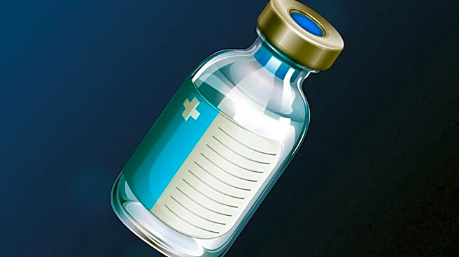
Beyond its primary anatomical and functional roles, the vastus lateralis muscle holds particular medical importance as a recommended site for intramuscular injections. This recommendation is specifically for infants less than 7 months old and for individuals who are unable to walk or who experience a loss of muscular tone. The muscle's large size and accessibility, combined with its distance from major nerves and blood vessels, make it a relatively safe and effective location for administering medications directly into the muscle tissue.
The use of the vastus lateralis for injections in these vulnerable populations minimizes the risk of injury to delicate structures and ensures proper absorption of the medication. Its robust nature and the volume of muscle tissue available make it an ideal choice when other injection sites might be less suitable or carry higher risks. This medical application underscores the practical significance of understanding the anatomy and characteristics of the vastus lateralis in clinical practice.