





Snapping hip syndrome, also known as dancer's hip, is a medical condition characterized by a distinct snapping sensation felt when the hip is flexed and extended. This sensation may be accompanied by an audible snapping or popping noise, and often, pain or discomfort. While the pain frequently diminishes with rest and reduced activity, the condition can persist for months or even years without appropriate treatment, sometimes becoming quite debilitating.
Understanding the different classifications and underlying causes is crucial for effective management.
Classifying Snapping Hip Syndrome: Intra-articular vs. Extra-articular
Snapping hip syndrome is commonly classified based on the location of the snapping: extra-articular or intra-articular. Intra-articular snapping hip syndrome often indicates an underlying mechanical problem within the hip joint itself. This can include injuries such as a torn acetabular labrum, ligamentum teres tears, loose bodies within the joint, articular cartilage damage, or synovial chondromatosis, which involves cartilage formations in the joint's synovial membrane. The pain associated with the intra-articular type tends to be more intense and debilitating compared to the extra-articular variety.
Extra-articular snapping hip syndrome, on the other hand, is frequently linked to factors outside the joint. These can include a leg length difference, typically with symptoms on the longer side, tightness in the iliotibial band (ITB), weakness in hip abductors and external rotators, poor lumbopelvic stability, and abnormal foot mechanics like overpronation. Popping in external snapping hip occurs when the thickened posterior aspect of the ITB or the anterior gluteus maximus rubs over the greater trochanter during hip extension. Internal snapping hip, a medial-extra articular type, is usually described as a snapping or locking of the hip with an audible sound, occurring when the iliopsoas tendon snaps over underlying bony prominences. Nearly half of patients with internal snapping hip also present with intra-articular pathology, highlighting the potential for overlapping issues.
Understanding the Causes and Risk Factors
The exact causes of snapping hip syndrome are not fully understood, but the onset is often insidious, beginning with a non-painful sensation or audible snapping during certain activities. Individuals may initially ignore this sensation, which can unfortunately lead to future discomfort. Athletes face an increased risk due to repetitive and physically demanding movements. This includes ballet dancers, gymnasts, horse riders, track and field athletes, soccer players, and individuals undergoing military training or engaging in vigorous exercise. Repeated hip flexion in these activities can lead to injury. In cases of excessive weightlifting or running, the condition is often attributed to extreme thickening of the tendons in the hip region. Snapping hip syndrome most commonly affects individuals between 15 and 40 years old.
Treatment Approaches: From Conservative to Medical Interventions
Fortunately, snapping hip syndrome is usually curable with appropriate treatment, and in some instances, it may even resolve spontaneously. If the condition is painless, there is generally little cause for concern. The primary goals of treatment are to correct any contributing biomechanical abnormalities and to stretch tightened muscles, such as the iliopsoas muscle or iliotibial band, to prevent recurrence. If self-treatment is unsuccessful or the injury interferes with daily activities, referral to a professional for an accurate diagnosis is necessary.
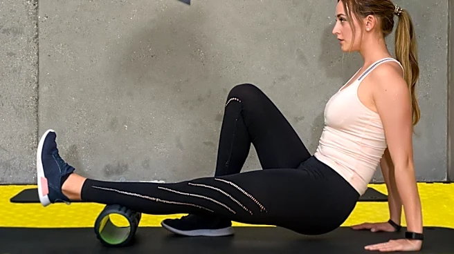
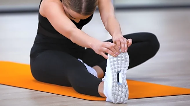
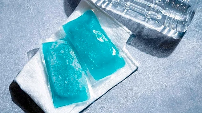
Medical treatment involves determining the underlying pathology and tailoring therapy accordingly. An examiner may assess muscle-tendon length and strength, perform joint mobility testing, and palpate the affected hip over the greater trochanter during activities like walking to identify lateral symptoms. Self-treatment often includes a HI-RICE regimen (Hydration, Ibuprofen, Rest, Ice, Compression, Elevation) for at least 48 to 72 hours after pain onset, particularly for soft tissue injuries of the iliopsoas muscle. Rest entails avoiding activities like running, hiking, jumping jacks, sit-ups, or leg lifts. Stretching tight structures such as the piriformis, hip abductor, and hip flexor muscles can alleviate symptoms. This involves stretching the involved muscle for 30 seconds, repeated three times with 30-second to 1-minute rest periods, performed twice daily for six to eight weeks, allowing for a gradual return to activities like jogging.
Conservative measures, including active and passive stretching exercises focusing on hip and knee extension, can resolve the problem in six to eight weeks. Stretching the hip into extension and limiting excessive knee flexion helps maximize the stretch to the iliopsoas tendon by avoiding passive insufficiency of the rectus femoris. Strengthening exercises for the hip flexors may also be part of the program, alongside non-steroidal anti-inflammatory drugs and activity modification. Once symptoms decrease, a maintenance program of stretching and strengthening, preceded by light aerobic activity, is important for long-term prevention. Injection-based treatments, typically focused on the iliopsoas bursa, often involve corticosteroid injections, which provide relief for weeks to months but carry side effects like weight gain, tissue weakening, and osteoporosis with regular use. While cellular-based therapy may hold future promise, current research has not yet proven its effectiveness.