

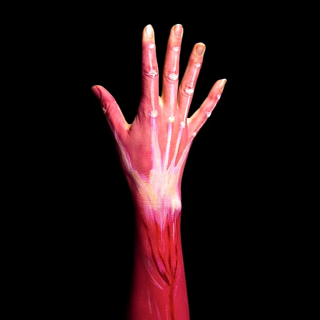


The pronator quadratus is a distinctive square-shaped muscle located in the distal forearm, playing a crucial role in the movement and stability of the hand and forearm. Its primary function is to pronate the hand, which means turning the palm downwards. This muscle is unique because it is the only one that directly connects the ulna and radius bones, attaching exclusively to these two bones at its respective ends. Understanding its structure, innervation,
and function provides insight into the intricate mechanics of the human forearm.
Anatomical Structure and Origin
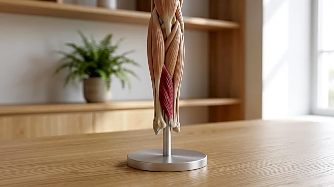
The pronator quadratus muscle is characterized by its square shape and its fibers run perpendicular to the length of the arm. It originates from the most distal quarter of the anterior ulna, which is the medial bone of the forearm. From this origin, its fibers extend to insert into the distal quarter of the radius, the lateral bone of the forearm. This specific arrangement allows it to effectively pull the radius across the ulna during pronation.
The muscle is composed of two distinct heads: a superficial head and a deep head. The superficial head originates from the anterior distal aspect of the diaphysis, or shaft, of the ulna. It then inserts into the anterior distal diaphysis of the radius, as well as its anterior metaphysis. The deep head shares the same origin point on the ulna but inserts slightly more proximally, closer to the ulnar notch of the radius. This dual-headed structure contributes to its efficiency in forearm movement.
Innervation, Blood Supply, and Function
The pronator quadratus muscle receives its nerve supply from the median nerve, specifically through its anterior interosseous nerve branch. This innervation is vital for the muscle's ability to contract and perform its function. Arterial blood supply to the pronator quadratus comes via the anterior interosseous artery, ensuring it receives the necessary nutrients and oxygen to operate effectively. This dedicated blood supply highlights its importance in forearm mechanics.
When the pronator quadratus contracts, its main action is to pull the lateral side of the radius towards the ulna. This movement results in the pronation of the hand, turning the palm to face downwards. Beyond its primary role in pronation, the deep fibers of the pronator quadratus also serve a critical function in maintaining the integrity of the forearm. They help to keep the ulna and radius, the two main bones of the forearm, bound together, contributing to the overall stability of the forearm joint. While it is a key pronator, it's worth noting that the pronator teres muscle also plays a major role in this action, and in some individuals, the pronator quadratus can even be absent without significantly affecting pronation.
Neurological Pathways for Control and Sensation
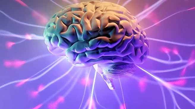
The motor pathway for the pronator quadratus is governed by the lateral corticospinal tract. This complex pathway begins in the precentral gyrus of the motor cortex, where a signal is initiated. This signal then travels from the upper motor nerve through the progression tracts of the internal capsule and continues through the cerebral peduncles of the midbrain. It then decussates, or crosses over, in the medulla and descends along the lateral corticospinal tract within the lateral column of the spinal cord. Finally, it decussates again in the spinal cord and synapses at the anterior horn with the lower motor neurons that control the skeletal muscles, including the pronator quadratus.
For sensation, the cuneate fasciculus tract is responsible for conveying information regarding the pronator quadratus's position and movement, as well as deep touch, visceral pain, and vibration. This sensory pathway starts in the dorsal nerve root, where the signal is transmitted through the dorsal horn and ascends the posterior column of the spinal cord. It synapses with an interneuron in the gracile nucleus, then decussates in the medial lemniscus of the medulla. The signal continues through the cuneate nucleus and the medial lemniscus of the midbrain before synapsing in the thalamus. A third-order neuron then transmits this signal to the postcentral gyrus of the somesthetic cortex, allowing for conscious perception of the muscle's state. These pathways are general to upper limb muscles but illustrate the sophisticated control and feedback mechanisms for the pronator quadratus.