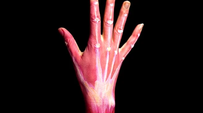





The adductor pollicis is a crucial muscle located in the hand, specifically responsible for the adduction of the thumb. This action involves bringing the thumb back into the plane of the palm from an abducted position, or moving it towards the side of the palm and index finger. Understanding its detailed anatomy, including its distinct heads, points of origin and insertion, and its innervation, provides insight into the precise mechanics of thumb movement
and its clinical significance in diagnosing certain nerve conditions.
Structural Components: Transverse and Oblique Heads
The adductor pollicis muscle is not a single, uniform structure but is composed of two distinct parts: the transverse head and the oblique head. These two heads originate from different points within the hand, contributing to the muscle's broad attachment and its ability to exert force across the thumb's carpometacarpal joint.
The transverse head, also known as the adductor transversus pollicis, is deeply seated. It originates from the anterior body of the third metacarpal bone. The oblique head, on the other hand, has a more complex origin, arising from the bases of the second and third metacarpals, as well as the adjacent trapezoid and capitate bones. This dual origin allows the adductor pollicis to effectively pull the thumb towards the palm.
Insertion and Innervation
Despite their separate origins, the fibers from both the transverse and oblique heads converge to a common insertion point. The greater number of fibers from the oblique head pass obliquely downward and unite with a tendon. This tendon, in turn, joins with the tendons of the medial portion of the flexor pollicis brevis and the transverse head of the adductor pollicis. The combined tendon then inserts into the ulnar side of the base of the proximal phalanx of the thumb. In individuals aged 14 or older, a sesamoid bone, often referred to as the adductor sesamoid, is present within this tendon.
Furthermore, a considerable fasciculus from the oblique head passes more obliquely beneath the tendon of the flexor pollicis longus. This fasciculus then joins the lateral portion of the flexor pollicis brevis and the abductor pollicis brevis. The adductor pollicis muscle receives its nerve supply from the deep branch of the ulnar nerve, specifically from spinal root T1. This innervation is vital for the muscle's function, and any compromise to the ulnar nerve can directly impact the adductor pollicis.
Actions and Clinical Relevance
The primary action of the adductor pollicis is to adduct the thumb at the carpometacarpal joint. While it is the main muscle responsible for this movement, other muscles such as the flexor pollicis brevis and the opponens pollicis also assist in thumb adduction. The ability to adduct the thumb is essential for many fine motor skills and grasping actions.
Clinically, the adductor pollicis muscle plays a significant role in diagnosing ulnar nerve damage. Froment's sign is a specific test used to assess for a compromised adductor pollicis muscle. During this test, a patient is asked to hold a piece of paper between their thumb and index finger. If the adductor pollicis is weak due to ulnar nerve dysfunction, the patient will compensate by flexing the interphalangeal joint of the thumb, indicating a positive Froment's sign. Additionally, in neuromuscular monitoring, the strength of adductor pollicis contraction is measured after stimulating the ulnar nerve, providing an objective assessment of nerve function.