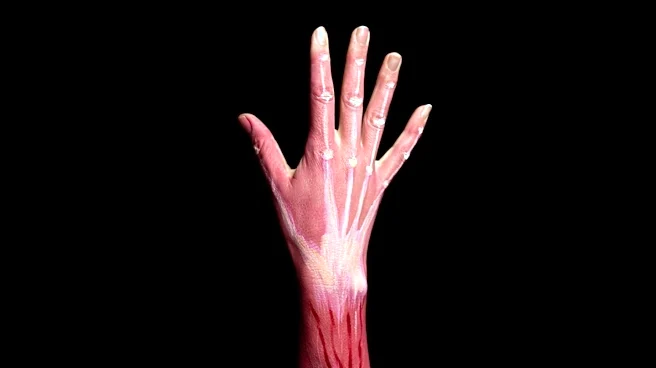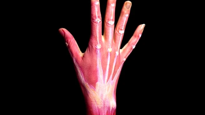









The pronator teres is a significant muscle located primarily in the forearm, playing a crucial role in the movement and stability of the arm. Alongside the pronator quadratus, its main function is to pronate the forearm, a motion that involves turning the palm to face posteriorly when the arm is in the anatomical position. This muscle's structure is complex, featuring two distinct heads, and its innervation by the median nerve highlights its importance
in upper limb mechanics. Understanding its anatomy, origin, insertion, and nerve supply is fundamental to comprehending forearm function.
Structural Components and Origins
The pronator teres muscle is characterized by its two heads: the humeral head and the ulnar head. The humeral head is the larger and more superficial of the two. It originates from the medial supracondylar ridge of the humerus, specifically from an area immediately superior to the medial epicondyle. This origin also includes fibers from the common flexor tendon, which itself arises from the medial epicondyle. This broad origin provides a strong anchor for the muscle's primary action.
In contrast, the ulnar head, sometimes referred to as the ulnar tuberosity, is a thinner bundle of muscle fibers. It originates from the medial side of the coronoid process of the ulna. Both the humeral and ulnar heads converge, joining at an acute angle, before the muscle continues its path across the forearm. This dual-headed structure is important because the median nerve, which supplies the muscle, passes between these two heads as it enters the forearm, separating it from the ulnar artery.
Insertion and Functional Role
After its origin, the pronator teres muscle extends obliquely across the forearm. It terminates in a flat tendon that inserts into a rough impression located at the middle of the lateral surface of the body of the radius. This insertion point is situated just distal to where the supinator muscle inserts. The specific placement of its insertion on the radius is key to its ability to pronate the forearm, effectively rotating the radius over the ulna.
The primary actions of the pronator teres are pronation of the forearm and flexion of the elbow. Pronation is the movement that turns the palm downward or backward. The muscle's cylindrical shape, reflected in its Latin name "teres" meaning "round or cylindrical shaped," is well-suited for this rotational movement. The word "pronator" itself comes from the Latin "pronus," meaning "inclined forward or lying face downward," directly referencing the muscle's action. In addition to its main role, the lateral border of the pronator teres forms the medial boundary of the cubital fossa, a triangular hollow found anterior to the elbow, further highlighting its anatomical significance.
Nerve Supply and Clinical Relevance
The pronator teres muscle receives its nerve supply from the median nerve, with contributions from nerve roots C6 and C7. The pathway for stimulating this muscle begins in the precentral gyrus of the brain, descending through the internal capsule and corticospinal tracts. After decussation at the medullary pyramids, the signal travels down the lateral corticospinal tract to the ventral horns of C5, C6, C7, C8, and T1. From there, it passes through the ventral rami and root ganglions of the brachial plexus, eventually reaching the median nerve branch that directly stimulates the pronator teres to contract, resulting in forearm pronation.
Beyond its normal function, the pronator teres is also implicated in a condition known as pronator teres syndrome. This is a compression neuropathy of the median nerve at the elbow, often occurring as the nerve passes between the two heads of the muscle. In certain clinical scenarios, such as C5 tetraplegia or radial nerve palsy, the pronator teres tendon can be rerouted in a procedure called tendon transfer. This rerouting, typically to the extensor carpi radialis brevis tendon, aims to restore wrist extension, demonstrating the muscle's versatility and importance in reconstructive surgery.