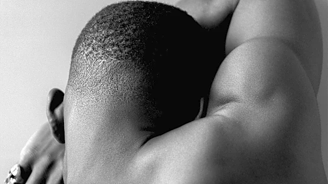

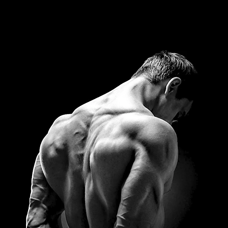





The trapezius is a large, paired surface muscle located in the back, historically known as the *cucullaris*. This muscle is distinctive for its trapezoid shape, which is how it gets its modern name. It extends significantly, originating from the occipital bone at the base of the skull and reaching down to the lower thoracic vertebrae of the spine. Laterally, it connects to the spine of the scapula, or shoulder blade. Its primary roles involve moving
the scapula and providing support for the arm, making it a crucial component of upper body movement and stability.
Structural Divisions and Origins
The trapezius muscle is not a single, uniform sheet but is divided into three distinct functional parts: superior (upper or descending) fibers, middle (transverse) fibers, and inferior (lower or ascending) fibers. Each section has specific origins and insertions, contributing to the muscle's overall broad reach and diverse functions. The superior fibers, for instance, originate from the spinous process of the seventh cervical vertebra (C7), the external occipital protuberance, the medial third of the superior nuchal line of the occipital bone, and the nuchal ligament. These fibers then extend downward and laterally to insert into the posterior border of the lateral third of the clavicle.
The middle fibers of the trapezius arise from the spinous process of the seventh cervical vertebra and the spinous processes of the first, second, and third thoracic vertebrae. They insert into the medial margin of the acromion and the superior lip of the posterior border of the spine of the scapula. The inferior fibers originate from the spinous processes of the remaining thoracic vertebrae, specifically from T4 through T12. These fibers proceed upward and laterally, converging near the scapula to end in an aponeurosis. This aponeurosis glides over a smooth triangular surface on the medial end of the spine, inserting into a tubercle at the apex of this surface. This intricate arrangement of fibers allows the trapezius to perform a variety of movements.
Connections and Fascial Relationships
At its occipital origin, the trapezius is connected to the bone by a thin, fibrous lamina that is firmly attached to the skin. The superficial and deep epimysia of the trapezius are continuous with an investing deep fascia. This fascia not only encircles the neck but also encloses both sternocleidomastoid muscles, highlighting the interconnectedness of the musculature in this region. In the middle section, the muscle connects to the spinous processes via a broad, semi-elliptical aponeurosis. This aponeurosis extends from the sixth cervical vertebra to the third thoracic vertebra and, along with the aponeurosis of the opposite trapezius muscle, forms a distinct tendinous ellipse known as the oval aponeurosis. The remaining parts of the muscle arise from numerous short tendinous fibers.
Functional Contributions and Clinical Relevance
The contraction of the trapezius muscle can produce two main effects: movement of the scapulae when the spinal origins are stable, and movement of the spine when the scapulae are stable. The upper part of the trapezius is responsible for supporting the weight of the arm. The middle region primarily retracts the scapula, pulling it closer to the spine. The lower part medially rotates and depresses the scapula. When all three fiber groups activate together, the upper and lower fibers assist the middle fibers, along with other muscles like the rhomboids, in scapular retraction or adduction. Dysfunction of the trapezius can lead to conditions such as winged scapula, sometimes specifically termed "lateral winging," and abnormal mobility or function of the scapula, known as scapular dyskinesia. Trapezius palsy, often resulting from damage to the spinal accessory nerve, can cause difficulty with arm adduction and abduction, a drooping shoulder, and pain in the shoulder and neck. The trapezius is also one of the muscles frequently affected in facioscapulohumeral muscular dystrophy (FSHD).