







Signs You Might Have a Fracture
While a sprain and a fracture can sometimes feel similar, certain symptoms are strong indicators of a broken bone. A sudden, sharp pain at the time of injury, especially if you heard a snapping or cracking sound, is a major red flag. Other tell-tale signs
include significant swelling and bruising that appears quickly, a visible deformity where a limb looks bent or out of place, and an inability to put weight on or use the injured area. Intense tenderness directly over the bone, rather than in the surrounding soft tissue, is another key symptom that should prompt a visit to a doctor.
How X-Rays See Inside Your Body
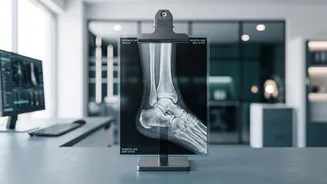
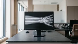
An X-ray is a fast and non-invasive imaging technique that provides a clear picture of your internal bone structure. The technology works by passing a small, safe amount of radiation through the body. Different tissues absorb this radiation differently. Dense structures like bone are rich in calcium and absorb most of the radiation, causing them to appear bright white on the final image. Less dense soft tissues, such as muscle and fat, allow more radiation to pass through and appear in shades of grey. A break or fracture disrupts the solid, white structure of the bone, showing up as a dark line on the image that a radiologist can identify.
What to Expect During the Procedure
Getting an X-ray is typically a quick and painless process, often taking just a few minutes. You will be asked to remove any clothing, jewellery, or metal objects that might interfere with the image. A technologist will position the injured part of your body between the X-ray machine and a detector plate. To get a complete picture, they will usually take at least two images from different angles, such as front-to-back and from the side. This is because some fractures can be hidden from a single viewpoint. You will need to stay very still for a few seconds while each image is taken to avoid blurring. You might also be given a lead apron to shield other parts of your body from radiation.
Reading the Results
Once the images are captured, a doctor or radiologist will examine them to determine the nature of the injury. They look for the location and type of fracture, such as a complete break, a partial crack, or a comminuted fracture where the bone is in several pieces. The X-ray also shows if the broken bone fragments are still aligned correctly or if they are displaced, which is crucial information for deciding on the best course of treatment—whether that involves a simple cast or requires surgery to realign the bone.
When an X-Ray Isn't Enough
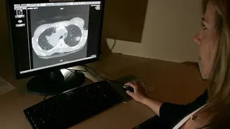
While X-rays are excellent for diagnosing most fractures, they do have limitations. They are not effective at showing soft tissue injuries like torn ligaments or muscle damage. Furthermore, certain types of fractures may not be visible on an initial X-ray, especially hairline or stress fractures, which are tiny cracks in the bone from repetitive force. Some fractures in complex joints like the wrist or hip can also be difficult to detect. If symptoms persist despite a normal-looking X-ray, your doctor may recommend a repeat scan in 7-10 days or use more advanced imaging like a CT scan or an MRI. A CT scan provides more detailed cross-sectional images of the bone, while an MRI is superior for visualising soft tissues and subtle bone injuries.