





The Skeleton Inside the Cell
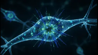
Just like our bodies rely on a bony skeleton for support, every cell in our body, including the billions of neurons in our brain, has its own internal scaffolding. This is known as the cytoskeleton. For decades, it was thought to be a relatively simple
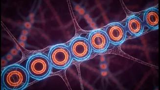
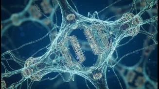
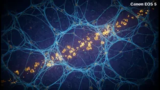
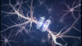
structure of protein filaments that helped the cell maintain its shape. However, recent discoveries have unveiled a far more intricate and dynamic structure just beneath the neuron's surface, known as the membrane-associated periodic skeleton (MPS). Instead of random filaments, scientists found that key proteins like actin form highly organised, repeating rings that wrap around the long axons of neurons, spaced at perfect intervals. This lattice-like structure was previously invisible, its details just below the resolution of older microscopes. Its discovery was a surprise, revealing a level of organisation that scientists hadn't predicted.
Seeing the Unseeable
Mapping this hidden skeleton presented a monumental challenge. The structures are incredibly small, with the repeating pattern spaced at about 190 nanometers—thousands of times thinner than a human hair. Traditional light microscopy wasn't powerful enough to resolve these details. The breakthrough came from the use of super-resolution microscopy and cryo-electron tomography (cryo-ET). These revolutionary techniques allow scientists to see the inner workings of a cell with near-atomic precision. Cryo-ET involves flash-freezing the cells, preserving their components in a near-natural state. Researchers can then use powerful electron microscopes to take a series of images from different angles, which a computer then reconstructs into a detailed 3D model. This process has finally allowed them to visualise the precise, organised rings of the MPS.
More Than Just a Scaffold
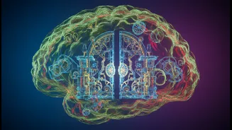
The new maps are revealing that the MPS is much more than a passive support structure. Recent findings suggest it acts as a crucial 'gatekeeper' for the neuron. Brain cells are constantly taking in materials from their surroundings, a process called endocytosis, which is vital for learning, memory, and general maintenance. Research from Penn State has shown that the MPS actively controls this process, regulating where and when substances can enter the cell. It acts as a physical barrier, with 'gates' that can open to allow nutrients in when needed. This regulatory role is a major shift in our understanding of how neurons function on a minute-to-minute basis.
A New Map for Neurological Diseases
The discovery and mapping of this internal skeleton have profound implications for understanding neurodegenerative diseases like Alzheimer's. Scientists found that when the MPS is weakened, it allows for an accelerated uptake of harmful proteins, such as those linked to Alzheimer's disease. This creates a destructive feedback loop: increased uptake further damages the skeleton, allowing even more harmful material to enter. This suggests that the integrity of this internal skeleton is a crucial line of defence for the brain. A breakdown in this cytoskeletal structure has also been linked to other conditions, including ALS and Charcot-Marie-Tooth disease. By understanding how the MPS is built and maintained, researchers hope to find new ways to protect it, potentially slowing or preventing the cellular damage that leads to these devastating conditions.