








The Challenge: Seeing Life’s Smallest Machines
Cryo-electron microscopy (cryo-EM) has been a game-changer in structural biology, even earning a Nobel Prize in 2017 for its ability to reveal the atomic-level details of life's molecular machinery. The technique involves flash-freezing biological molecules
in a thin layer of ice and then imaging them with an electron beam. This allows scientists to determine the three-dimensional structures of proteins, viruses, and other cellular components without having to crystallise them. However, cryo-EM has a significant blind spot: small proteins. Proteins smaller than about 70 kilodaltons—which constitute an estimated 90% of all proteins in the human body—have remained stubbornly difficult to image clearly. This is because they generate a very weak signal, resulting in low-contrast, 'washed-out' images that are difficult to analyse. This limitation has left a massive gap in our understanding of human health and disease, as these small proteins are often crucial drug targets.
A Leap Forward: The Laser Phase Plate
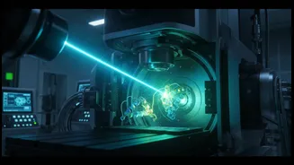
Scientists at the University of California, Berkeley, and Biohub have pioneered a solution that adapts a century-old optical technique for the modern era of electron microscopy. Their invention, the laser phase plate, dramatically boosts the contrast of cryo-EM images. The concept is rooted in phase-contrast microscopy, which won a Nobel Prize in 1953 for allowing biologists to see faint structures inside cells under a light microscope. Applying this to an electron microscope, which has 10,000 times the magnification, required a monumental engineering feat. It involves building an apparatus with an intensely powerful, focused continuous-wave laser—brighter than the sun—that interacts with the electron beam after it passes through the sample. This interaction shifts the phase of the electrons, turning previously invisible details into sharp, high-contrast features.
How It Sharpens the Picture
In conventional cryo-EM, contrast is typically generated by slightly defocusing the microscope, but this compromises the quality and resolution of the final image. The laser phase plate solves this problem by directly manipulating the electron beam itself. By trapping laser light between two perfectly polished mirrors, the system creates an intense electromagnetic field that shifts the phase of the unscattered electrons by 90 degrees. This phase shift dramatically increases the contrast of the image without sacrificing resolution. The Berkeley team demonstrated the power of this new system, which they named Theia, by imaging hemoglobin—a protein on the very edge of what is possible with today's standard cryo-EM machines. The results were a significant improvement, providing a much clearer and more detailed structure.
Unlocking New Frontiers in Medicine
The ability to see small proteins clearly is a massive step forward for drug discovery and disease research. A huge number of human proteins involved in conditions like cancer, neurodegenerative diseases, and metabolic disorders are smaller than the previous imaging threshold. This technology allows researchers to visualise these once-elusive molecules, understand their function, and identify new sites for drugs to bind. It is particularly revolutionary for a technique called cryo-electron tomography (cryo-ET), which creates 3D images of molecules within their natural cellular environment. This provides context, showing how proteins interact with each other inside a crowded cell—a bit like going from seeing a single tree to understanding the entire forest.
What This Means for India’s Research Ecosystem
For the burgeoning biotechnology and pharmaceutical research sectors in India, this advancement is incredibly significant. Leading Indian institutions like the Indian Institute of Science (IISc) in Bengaluru and the National Centre for Biological Sciences (NCBS) have already invested heavily in state-of-the-art cryo-EM facilities. This new laser phase plate technology, once commercialised and integrated, could supercharge their research capabilities. It would empower Indian scientists to tackle structural biology challenges that were previously out of reach, accelerating the design of novel therapeutics for diseases pertinent to the Indian population. The ability to perform structure-based drug design on a wider array of protein targets can make drug discovery pipelines more efficient and innovative, positioning India to be a leader in a new era of molecular medicine.