





The Challenge of Seeing Proteins
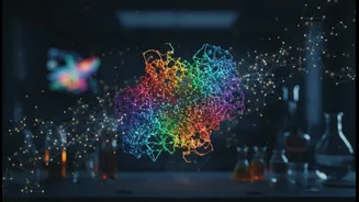
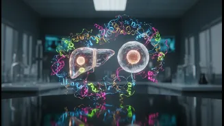
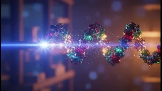
Proteins are the workhorses of our bodies. They are microscopic machines that carry oxygen, build tissues, send signals, and fight off diseases. Understanding their exact three-dimensional shape is the key to understanding how they work—and how they malfunction
in diseases like cancer or Alzheimer's. For decades, this has been a central goal of biology. But there's a problem: most proteins are incredibly small and essentially transparent. Visualizing them is like trying to take a crystal-clear photo of a tiny, invisible object. For years, scientists have used a revolutionary technique called cryo-electron microscopy (cryo-EM), which freezes molecules and uses electrons to image them. While cryo-EM has been a game-changer, it has struggled with the smallest and most delicate proteins, which make up the majority of those in the human body. The images often lack contrast, making it difficult to determine the precise structure needed for designing targeted drugs.
A Breakthrough of Light and Electrons
Now, a device called a laser phase plate, developed by researchers at UC Berkeley and the Chan Zuckerberg Biohub, is changing the game. The concept is an elegant solution to the contrast problem. In traditional microscopy, contrast is often generated by slightly defocusing the image, but this sacrifices sharpness and loses information. A phase plate is a tool that can boost contrast without this trade-off. However, previous physical phase plates suffered from issues like charging and degradation under the intense electron beam. The new approach is completely different. Instead of a physical object, it uses an intensely powerful laser beam focused to a microscopic point. The electron beam of the microscope passes through this laser field, which subtly alters the phase of the electrons, dramatically increasing image contrast without the negative side effects of older methods.
How Does a Laser Phase Plate Work?
Think of it like adjusting the lighting in a photo studio. If your subject is washed out, a skilled photographer doesn't just turn up the main light; they add subtle side lights to create shadows and definition. The laser phase plate does something similar for electrons. The core idea, first proposed over a decade ago but long considered too difficult to build, involves creating a standing wave of light by bouncing a laser between two highly reflective mirrors thousands of time. This amplifies the laser's intensity to a staggering degree—hotter than the sun's surface—creating a powerful electromagnetic field. When the microscope's unscattered electrons pass through this field, their phase is shifted by exactly the right amount to create high-contrast interference with the electrons that were scattered by the protein sample. The result is that previously faint or invisible molecular details pop into sharp relief, turning a blurry mess into a clear blueprint.
Unlocking New Frontiers in Drug Discovery
So, why does a clearer picture of a protein matter so much? The answer is central to modern medicine. Designing a new drug is like creating a key for a specific lock. The 'lock' is the active site on a protein that is involved in a disease. If you don't know the exact shape of the lock, you can't design an effective key. By providing high-resolution images of proteins that were previously impossible to see clearly, the laser phase plate allows researchers to map these 'locks' with unprecedented accuracy. This is especially crucial for developing drugs for notoriously difficult targets, such as the small proteins that drive many cancers or neurodegenerative diseases. Furthermore, this technology enhances a technique called cryo-electron tomography (cryo-ET), which creates 3D images of proteins in their natural environment inside a cell, not just in isolation. This gives scientists a view of how these molecular machines actually operate and interact in the crowded, complex world of a living cell.
The Future of Seeing the Unseen
This technology represents a major leap forward, not just an incremental improvement. Scientists estimate that while current cryo-EM can effectively image perhaps 10% of the human proteome (the full set of our proteins), the laser phase plate could push that number above 50%. This opens the door to understanding vast, unexplored territories of human biology. Researchers are already using the technology to get better images of benchmark molecules like hemoglobin. The next step is to turn this powerful new microscope on the most complex and medically relevant targets. The ability to see what was once invisible promises to accelerate the pace of discovery, leading to more precise drugs with fewer side effects and offering new hope for treating some of our most challenging diseases.