





The Brain's Electrical Buzz
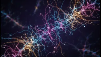
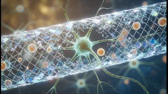
Imagine your brain as a dense network of billions of tiny wires called neurons. They communicate with each other at junctions called synapses. For a message to pass from one neuron to the next, it travels as an electrical pulse, known as an action potential,
down the length of the cell. But when this pulse reaches the end of the neuron, it needs help to cross the gap to the next one. This is where calcium comes in, acting as the critical link that turns an electrical signal into a chemical one, allowing communication to continue.
Calcium: The Spark of Communication
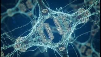
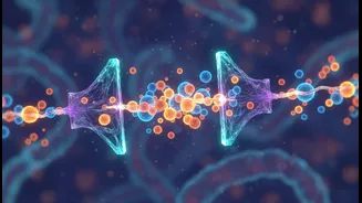
When the electrical signal arrives at the neuron's terminal, it triggers the opening of special doorways called voltage-gated calcium channels. As these channels open, calcium ions rush into the cell. This influx of calcium is the direct trigger that causes vesicles—tiny sacs filled with chemical messengers called neurotransmitters—to fuse with the cell membrane and release their contents into the synapse. These neurotransmitters then travel to the next neuron, delivering the message. Without this calcium spark, the signal would stop, and communication between brain cells would fail. It’s a process that happens in microseconds, fundamental to everything your brain does.
Meet the Gatekeepers: Calmodulin
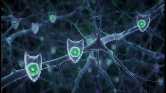
The flow of calcium cannot be an uncontrolled flood; it must be precisely regulated. Too much calcium can be toxic to cells. This is where the microscopic gatekeepers come into play. A key player in this process is a protein called calmodulin (CaM). This ancient and versatile protein acts as a calcium sensor. It is often physically associated with the calcium channels themselves. When calcium ions enter the cell, they bind to calmodulin. This binding causes calmodulin to change shape, which in turn influences the calcium channel's ability to stay open or to close, a process sometimes called calmodulation. This feedback loop ensures that calcium levels are perfectly managed, allowing just enough to enter to do the job without causing harm.
A Delicate Balancing Act
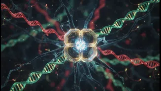
The regulation by proteins like calmodulin is a sophisticated dance. It can lead to two opposing effects: Ca2+-dependent inactivation (CDI) and Ca2+-dependent facilitation (CDF). In CDI, the binding of calcium to calmodulin helps to close the channel, preventing excessive calcium influx—a crucial protective mechanism. In CDF, under certain conditions, the calcium-calmodulin complex can actually make it easier for the channel to open, enhancing the signal. How a single protein can perform these opposing roles is a subject of intense research. This dual capability allows the brain to fine-tune its signals with incredible precision, strengthening some connections while dampening others, which is the basis for learning and memory.
When the Gates Fail
The importance of these gatekeepers becomes devastatingly clear when they malfunction. Disrupted calcium signaling is a feature of many neurological and psychiatric disorders. For instance, in conditions like Alzheimer's disease, evidence suggests that the regulation of calcium homeostasis is abnormal, which may contribute to neuron dysfunction and death. Similarly, in Parkinson's disease, alterations in certain calcium channels are thought to be involved in the vulnerability of dopamine-producing neurons. Growing evidence links problems with calcium influx and its regulation to a wide range of conditions, from epilepsy to schizophrenia, highlighting why understanding these microscopic mechanisms is a major frontier in neuroscience.