




Mapping the Brain's Command Centre
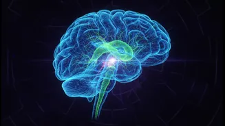
The brainstem is one of the most vital and complex regions of the human body. Connecting the brain to the spinal cord, it acts as a critical command centre, regulating essential functions we perform without a second thought: breathing, heart rate, sleep,
and balance. Given its importance, any damage or abnormality in the brainstem can have severe, often life-threatening consequences. For decades, scientists have sought to create detailed maps, or atlases, of this region to better understand its intricate structure. These atlases serve as a crucial reference, much like a geographical map, allowing researchers to navigate the brain's complex terrain and identify where problems occur.
The Need for an Indian-Specific Model
Until recently, neuroscientists in India largely relied on brain atlases created from Caucasian brains, most notably the Montreal Neurological Index (MNI) template. However, research has shown that there are distinct differences in the size, shape, and structure of brains across different ethnic populations. Studies have revealed that the average Indian brain is smaller in height, width, and volume compared to its Caucasian counterparts. When a standard MRI scan from an Indian patient is compared against a Caucasian brain template, these anatomical differences can be misinterpreted as abnormalities, potentially leading to misdiagnosis or inaccurate research findings. Recognizing this critical gap, Indian scientific institutions have been working to develop brain templates that are representative of the nation's population.
Introducing ANCHOR: A Groundbreaking Atlas
The latest breakthrough comes from the Sudha Gopalakrishnan Brain Centre at IIT Madras. Researchers there have developed what is being described as the world's most detailed 3D atlas of the human brainstem, named ANCHOR (Atlas of Neurochemical Characterization of the Human Brainstem with 3D Reconstruction). What makes ANCHOR exceptional is its incredible detail. Scientists painstakingly reconstructed maps of over 200 different nuclei and fibre tracts from hundreds of tissue sections, using advanced imaging and multiple chemical stains to identify various cell types. This provides a multi-layered view, allowing a user to zoom from a broad MRI view right down to the cellular level. Furthermore, the project includes data from prenatal, childhood, and adult brains, offering an unprecedented look at how this critical structure develops over a human lifespan.
A New Weapon Against Neurological Disease
The implications of this detailed atlas are immense for medical research. It provides a powerful tool to study conditions where the brainstem is affected, such as Parkinson's disease, multiple system atrophy, Alzheimer's, dementia, and strokes. By having a precise, high-resolution map, scientists can more accurately pinpoint the specific groups of cells that are damaged by a particular disease. This could lead to earlier and more accurate diagnoses, as well as the development of more targeted therapies. Ajay Kumar Sood, the Principal Scientific Adviser to the Government of India, noted that these maps will be critical for clinical applications by helping to identify the exact cell populations affected in brainstem lesions.
Free Access to Fuel Global Collaboration
Perhaps one of the most significant aspects of the ANCHOR atlas is that it has been made publicly and freely available to researchers and clinicians worldwide. This open-access approach is a deliberate effort to democratise science and accelerate progress. By removing financial barriers, the team at IIT Madras has ensured that any researcher, from a large university to a smaller lab, can use this state-of-the-art tool. This fosters a collaborative environment, allowing scientists globally to pool data and build upon each other's work more effectively. This initiative is part of a broader programme at the Sudha Gopalakrishnan Brain Centre to create a comprehensive repository of human brain maps, a crucial step in decoding the complexities of the human brain and its disorders.