






The Brain's Inner Scaffolding
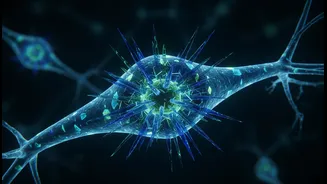
Every cell in our body, including the incredibly complex neurons in our brain, has an internal skeleton known as the cytoskeleton. It’s a network of protein filaments that provides shape, structure, and a transport system for cellular components. For
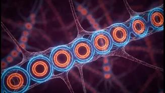
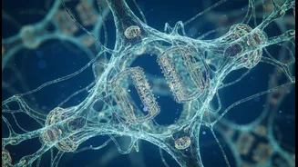
a long time, one part of this structure in neurons—a lattice just beneath the cell's surface called the membrane-associated periodic skeleton, or MPS—was considered to be nothing more than structural support. Discovered in 2013, this intricate mesh of actin and spectrin proteins was believed to simply help the long, cable-like axons of neurons maintain their form. It was an important, but seemingly boring, part of the cell's architecture. However, new research from Penn State has completely upended this view, revealing a function far more critical than just holding the cell together.
From Passive Frame to Active Gatekeeper
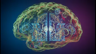
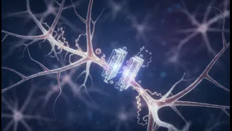
The recent study, published in the journal Science Advances, shows that the MPS is not a passive frame but an active and vital gatekeeper. Researchers found it dynamically controls a fundamental process called endocytosis—the method by which neurons absorb substances from the fluid surrounding them. This includes essential nutrients and signaling molecules needed for learning, memory, and the daily upkeep of the brain. The MPS acts as a physical barrier, essentially putting the brakes on how quickly the neuron can pull things inside. When researchers artificially disrupted this skeleton, they observed that the cells began to absorb materials at a much faster rate, confirming that the MPS normally slows this process down. This discovery transforms our understanding of the MPS from a simple scaffold into a sophisticated regulator of cellular traffic.
How Scientists Saw the Invisible
Observing this function required peering into a world far too small for conventional microscopes. The components of the MPS are arranged in a periodic pattern with a spacing of about 190 nanometres—well below the diffraction limit of visible light. To see it in action, the Penn State team, led by researcher Ruobo Zhou, used a powerful technique called super-resolution microscopy. This advanced imaging method allows scientists to visualize structures at the nanoscale, effectively creating a movie of molecular processes as they happen inside living cells. By tagging specific proteins with fluorescent markers, they could track how different molecules entered cultured neurons and directly observe the MPS regulating the flow. This technology was not only key to this new discovery but was also instrumental in the original identification of the MPS structure back in 2013.
A New Battleground for Alzheimer's
The most profound implication of this discovery is its connection to neurodegenerative diseases like Alzheimer's. One of the hallmarks of the disease is the buildup of toxic protein fragments, specifically amyloid-beta. These fragments are created when a larger protein, called amyloid precursor protein (APP), is absorbed by the neuron and broken down. The new research shows that when the MPS gatekeeper is weak or damaged—a condition observed in aging and neurodegenerative disease—the neuron absorbs APP much more rapidly. This creates a destructive feedback loop: increased absorption of APP leads to more toxic amyloid-beta, which can further damage the cell, including the MPS itself, leading to an even faster, uncontrolled intake. This suggests that the breakdown of this cellular skeleton could be a key event that pushes a neuron into a state of decline.
The Future of Brain Health
This new understanding of the MPS as a protective gatekeeper opens an entirely new front in the fight against neurodegeneration. Instead of only focusing on clearing out toxic proteins after they have formed, researchers can now explore strategies to protect and stabilize the MPS itself. The Penn State team suggests that preserving the integrity of this lattice could become a powerful therapeutic strategy to slow the progression of Alzheimer's. By strengthening the gate, it might be possible to prevent the neuron from taking in harmful amounts of proteins in the first place, thus preventing the toxic cascade before it even begins. While this research is still in its early stages, it represents a fundamental shift in how we view the inner workings of our most vital cells and offers a hopeful new direction for future treatments.