
The Challenge of Seeing the Invisible
Understanding disease and designing effective drugs often comes down to seeing the three-dimensional shape of proteins. These molecules are the workhorses of our cells, but many are incredibly small and delicate. For years, the gold standard for imaging
them was X-ray crystallography, which requires forcing proteins into a crystal form—something that is not always possible. Then came cryo-electron microscopy (cryo-EM), a Nobel Prize-winning technique where samples are flash-frozen and imaged with electrons. This method allowed scientists to see proteins in a more natural state. However, cryo-EM has its own limitations. To avoid destroying the fragile samples, a low-intensity electron beam is used, resulting in low-contrast, noisy images, especially for smaller proteins. This is like trying to take a photo in a dimly lit room; the resulting picture is often blurry and lacks detail, making it difficult to analyze over 90% of the proteins in human cells.
A Leap in Microscopic Vision
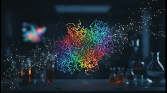
Scientists at UC Berkeley and Biohub have developed a groundbreaking solution: the laser phase plate. This innovation adapts a nearly century-old optical trick called phase contrast for the high-powered world of electron microscopes. In simple terms, the device uses an intensely powerful laser—focused within the microscope—to interact with the electron beam after it passes through the sample. This interaction shifts the phase of the electrons, dramatically boosting the contrast of the final image without damaging the specimen. The result is a much clearer, higher-resolution picture. Early demonstrations show a massive improvement in clarity, turning faint, blurry shapes into detailed structures. Researchers have already used it to get better images of proteins like hemoglobin, which was previously at the very edge of what cryo-EM could resolve.
From Blurry Pictures to Drug Blueprints
The ability to clearly see smaller proteins is a game-changer for drug discovery. Many diseases are caused by malfunctioning proteins, and drugs are often designed to fit onto these proteins like a key into a lock, altering their function. Without a precise blueprint of the 'lock,' designing an effective 'key' is a process of trial and error. The laser phase plate provides that blueprint with unprecedented detail. This clarity allows researchers to identify previously hidden pockets and surfaces on proteins that could be targeted by new medicines. Furthermore, the technology enhances a related technique called cryo-electron tomography (cryo-ET), which creates 3D images of proteins interacting inside their natural, crowded cellular environment. This provides critical context, showing how proteins function—and malfunction—in real-time, which is invaluable for developing therapies for complex illnesses.
Why Caution Is Still the Watchword
Despite the excitement, experts emphasize that the laser phase plate is still an emerging tool. The technology itself is an incredible feat of engineering, requiring mirrors polished to atomic-level smoothness and lasers that are exceptionally powerful and stable. These systems are not yet widely available and are currently installed in only a few custom-built, state-of-the-art microscopes. Researchers note that for some larger, easier-to-image proteins, the advantage of the laser is minimal. Its true power shines when dealing with the most challenging samples: very small proteins or messy, complex cellular environments. While initial results are transformative, the technology needs further refinement to become a standard, accessible tool for labs worldwide. It is a major step toward a new era of structural biology, but the journey has just begun.