






An Icon of Extinction
For centuries, the dodo has been a symbol of foolishness and obsolescence. Living on the island of Mauritius, isolated from natural predators, these flightless birds were unafraid of the European sailors who arrived in the late 16th century. This lack
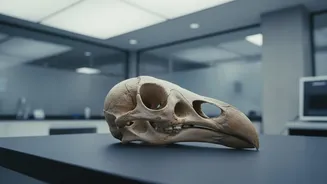
of fear proved fatal. Less than a hundred years after its discovery, the dodo was extinct, hunted and outcompeted by invasive species. Because their disappearance happened so quickly, very few complete skeletons remain, making each fossil incredibly precious. For a long time, our image of the dodo was based on sailors' exaggerated tales and a few fragmented bones. But what if we could look inside its skull to learn how it truly lived?
A Hospital for Fossils
Enter the world of non-destructive research. Paleontologists face a constant dilemma: to understand a fossil, you often need to cut it open, which destroys the very thing you're trying to study. This is where Computed Tomography (CT) scanning comes in. Just like a doctor uses a CT scanner to see inside a human body without surgery, scientists can use high-resolution scanners to peer inside priceless fossils. By taking thousands of X-ray cross-sections from every angle, a powerful computer can assemble a detailed, 3D digital model of the object. For the dodo, this technology is a game-changer. Researchers can now study the internal structure of the world's only two complete dodo skulls—one in Denmark, the other at Oxford—without risking even a single scratch.
Rebuilding a Virtual Brain
Once a dodo skull is scanned, the real detective work begins. Scientists use specialised software to create a digital endocast—a model of the cavity where the brain once sat. As a brain grows, it leaves impressions on the inside of the skull. These imprints are a fossilised map of the brain's shape and size, offering vital clues about the animal's senses and cognitive abilities. By analysing this virtual brain, researchers can estimate the size of different regions. For example, a large olfactory bulb would suggest a strong sense of smell, while the size of the optic lobes relates to vision. This process allows scientists to move beyond speculation and make evidence-based interpretations of the dodo's biology.
Answering the Science Questions
So, what have these digital reconstructions revealed? The findings are overturning the myth of the "dumb dodo." The scans show that the dodo's brain-to-body size ratio was comparable to that of modern pigeons, birds known for their intelligence and navigational skills. The most surprising discoveries relate to its senses. Contrary to what its small optic lobes might suggest, the dodo wasn't visually impaired but likely adapted for a different kind of sight. Evidence points to an animal that may have been crepuscular, meaning it was active during the low-light hours of dawn and dusk, possibly to avoid competing for food with giant tortoises. Furthermore, the dodo had a significantly larger olfactory bulb than its pigeon relatives, indicating a powerful sense of smell. Its large beak was also full of nerves, suggesting it was used as a sensitive tool to feel and explore its surroundings.
Beyond the Dodo
The non-destructive techniques pioneered in studies like this are revolutionizing paleontology. Scientists are applying the same methods to everything from dinosaur eggs to the fossilised remains of early humans and other extinct Australian fauna like the Tasmanian tiger. These digital fossils can be shared instantly with researchers across the globe, allowing for unprecedented collaboration. They serve as a permanent, high-fidelity archive of specimens that might be too fragile to handle or locked away in museum collections. This virtual approach ensures that future generations of scientists, armed with new questions and even more advanced technology, can continue to learn from the past without erasing it.