



The Challenge of Seeing Life’s Building Blocks
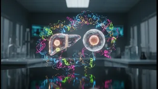
Proteins are the microscopic machines that drive nearly every process inside our bodies. Understanding their unique 3D shapes is critical for developing new medicines for everything from cancer to neurodegenerative diseases. For years, one of the most
powerful tools for this has been cryo-electron microscopy (cryo-EM). This technique freezes proteins and uses an electron beam to take their picture, a method so revolutionary it won a Nobel Prize. However, cryo-EM has a significant blind spot: it struggles to get clear, high-contrast images of smaller proteins. Since a majority of human proteins fall into this 'too small to see clearly' category, a huge part of our own biology has remained blurry, limiting the ability of researchers to design effective drugs.
A Breakthrough Forged in Light
Researchers at the University of California, Berkeley, and the Chan Zuckerberg Biohub have developed a game-changing solution that was once considered nearly impossible. They have created a 'laser phase plate'—an add-on for cryo-EM microscopes that dramatically boosts image contrast. The concept is an advanced adaptation of a phase-contrast technique that revolutionized light microscopes nearly a century ago. In simple terms, the device uses an incredibly intense, focused laser—100 million times brighter than the sun's surface—to interact with the microscope's electron beam. This interaction subtly changes the phase of the electrons, which in turn makes previously faint and washed-out parts of a protein structure appear in sharp, brilliant contrast without damaging the delicate sample.
From Blurry to Brilliant Detail
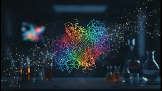
The results are stunning. In tests, the laser phase plate has enabled microscopes to produce clear images of proteins that were at the very limit of what was previously possible. For example, the structure of hemoglobin, the protein that carries oxygen in our blood, can now be seen with far greater detail. This breakthrough effectively lowers the size limit for what can be studied, opening up an estimated 50% of the human proteome to high-resolution imaging, compared to just 10% with older methods. This is particularly crucial for a related technique called cryo-electron tomography (cryo-ET), which aims to map proteins within their natural, crowded environment inside a cell. The improved contrast will help scientists untangle this complex scene, much like finding a specific leaf on a tree in a dense forest.
Accelerating the Future of Medicine
The implications for drug discovery are immense. If a protein is the 'lock' that a disease affects, a drug is the 'key' designed to fit it. The sharper the image of the lock, the more precisely scientists can design the key. By making tens of thousands of previously invisible proteins visible, this technology could unveil a vast new landscape of potential drug targets. This could accelerate research into treatments for today’s most challenging diseases. While AI tools like AlphaFold have made incredible strides in predicting protein structures, they rely on experimental data for validation. The laser phase plate provides exactly the kind of high-quality experimental data needed to confirm and refine these computational models, creating a powerful synergy between AI and physical imaging.