






The Limits of a Visual Exam
A physical examination is always the first step. A doctor will check for swelling, tenderness, bruising, and deformity. However, what you see on the surface doesn't tell the whole story. Many fractures, like small hairline cracks or stress fractures from
repetitive activity, may not cause dramatic visible signs. Swelling can also mask the true extent of an injury. What might feel like a severe sprain could actually be a fracture that, if left untreated, could lead to serious complications. Relying only on a visual check is often guesswork; medical imaging allows doctors to see beyond the surface symptoms for an accurate diagnosis.
X-Rays: The First Line of Defense
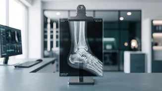
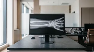
The X-ray is the gold standard and the most common first step in diagnosing a fracture. It uses a small amount of radiation to create images of your bones, which show up as bright white. A break or crack disrupts this solid white structure, appearing as a dark line. X-rays are fast, widely available, and highly effective at identifying a wide range of breaks, from clean breaks to more complex fractures where a bone is shattered into multiple pieces. This allows a clinician to confirm the fracture's location and basic pattern, which is essential for creating the right treatment plan, whether that involves a simple splint or a referral to a specialist.
When an X-Ray Isn't Enough
Sometimes, a fracture is so small or located in such a complex area that it doesn't show up on an initial X-ray. These are known as 'occult' or hidden fractures. They are common in certain areas like the wrist, hip, and ankle, as well as in cases of stress fractures. Because an X-ray is a 2D image of a 3D structure, a fracture line can sometimes be obscured by another bone. In these situations, even if you have clear symptoms of a fracture, the X-ray might look normal. Your doctor may then recommend a follow-up X-ray in 10-14 days, as the healing process can sometimes make the fracture more visible, or they may order more advanced imaging tests.
Advanced Imaging: CT and MRI Scans
When an X-ray is inconclusive or more detail is needed, doctors turn to Computed Tomography (CT) or Magnetic Resonance Imaging (MRI). A CT scan uses X-rays from multiple angles to create detailed, cross-sectional images of bone. It is excellent for assessing complex fractures, injuries to joints, or planning for surgery. An MRI, on the other hand, uses magnetic fields and radio waves to get a clear picture of soft tissues like ligaments, tendons, and cartilage, as well as bone. It is highly sensitive and can detect tiny stress fractures, bone bruises, or damage to the surrounding tissues that a CT scan or X-ray might miss.
Why an Accurate Diagnosis Matters
Getting a proper diagnosis is crucial because leaving a fracture untreated can lead to significant long-term problems. A bone that isn't properly aligned and immobilized may heal incorrectly (a condition called malunion) or fail to heal at all (nonunion). This can result in chronic pain, joint stiffness, reduced mobility, and an increased risk of arthritis in the future. In some cases, an untreated fracture can also damage surrounding nerves and blood vessels or lead to serious infections. By using the right imaging tools, doctors can ensure you receive the appropriate care, promoting proper healing and preventing these potentially serious complications.