








A Groundbreaking Achievement in Neuroscience
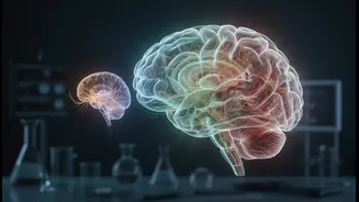
Researchers at the Indian Institute of Technology Madras have developed what is being called the world's most detailed 3D atlas of the human brainstem. Dubbed ANCHOR, which stands for Atlas of Neurochemical Characterization of the Human Brainstem with
3D Reconstruction, this digital map provides a view of the brainstem at cellular-level resolution. Developed by the Sudha Gopalakrishnan Brain Centre (SGBC), the atlas integrates multiple types of imaging, including MRI and detailed chemical staining of tissue, to create the most comprehensive map of its kind. Unlike previous maps, which were often two-dimensional or lacked fine detail, ANCHOR allows scientists to zoom from a full view of the brain down to the level of individual cells across different life stages.
Why the Brainstem Is So Critical
The brainstem is a small, stalk-like structure that connects the main part of the brain to the spinal cord. Despite its size, it's the command centre for most of our body's essential, unconscious functions. It regulates breathing, heart rate, blood pressure, sleep cycles, and balance. Because it controls these vital processes, even minor damage to the brainstem can have severe or life-threatening consequences. It contains more than 200 tiny, densely packed clusters of cells and nerve fibres, known as nuclei and tracts, making it incredibly complex to study. This new atlas provides a clear guide to this intricate territory for the first time.
Charting Growth from Foetus to Adult
One of the most significant features of the ANCHOR atlas is its developmental timeline. For the first time, researchers can observe how the brainstem's structure and chemical makeup change from the prenatal stage all the way through adulthood. This provides an invaluable window into how this critical part of the nervous system is built. By understanding the normal developmental trajectory, scientists can better identify when and where things might go wrong. This is crucial for studying developmental disorders that may have their origins in the earliest stages of life, offering a baseline against which abnormalities can be compared.
Unlocking Medical Mysteries
The potential applications for this high-resolution map are vast. Experts believe it could transform how neurological diseases are diagnosed and treated. By providing a precise reference, doctors may be able to pinpoint the specific groups of cells affected by conditions like Parkinson's disease, Alzheimer's, and other forms of dementia. For example, the atlas could help researchers create more accurate lab-grown models of the specific neurons that are damaged in Parkinson's, accelerating the development of cell-based therapies. Furthermore, it may shed light on conditions linked to brainstem abnormalities, such as certain sleep disorders and potentially even Sudden Infant Death Syndrome (SIDS), which has long been theorized to involve the brainstem's control over breathing and waking.
A Global Tool for Science
The team at IIT Madras has made the ANCHOR atlas publicly available, allowing scientists, doctors, and students around the world to benefit from this powerful new resource. This open-access approach is expected to accelerate global research efforts in neuroscience. Ajay Kumar Sood, the Principal Scientific Advisor to the Government of India, praised the project as a step towards decoding the “most complex creation” and a landmark achievement for Indian science. The atlas serves as a foundational tool that can be integrated with other data, helping researchers interpret clinical neuroimaging and improve the precision of brain surgery and other targeted treatments.