





The Brain’s Vital Command Centre
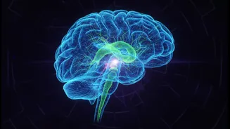
Think of the brainstem as the silent, tireless manager of your body. It’s the stalk-like structure connecting your brain to your spinal cord, and it governs the fundamental processes that keep you alive: breathing, heart rate, sleep-wake cycles, and consciousness
itself. For decades, this densely packed and incredibly complex region has been like a black box for neuroscientists. Its intricate network of cells and fibres was too dense to map with existing technology, leaving our understanding of its role in both health and disease frustratingly incomplete. Damage to this small area can have catastrophic consequences, yet doctors have often worked with vague maps, unable to see precisely which cells were affected by a stroke, tumour, or degenerative disease.
A Landmark Achievement by IIT Madras
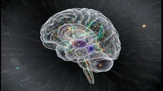
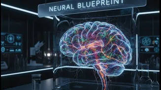
In a major leap forward for global neuroscience, a team led by researchers at the Indian Institute of Technology Madras (IIT Madras) has created the world's most detailed three-dimensional atlas of the human brainstem. Developed by the institute's Sudha Gopalakrishnan Brain Centre (SGBC), this map charts the brainstem's terrain down to the level of individual cells. Unveiled at the 3rd BRICS Neuroscience Symposium 2026, the project provides an unprecedented view of this crucial brain region, mapping over 200 distinct nuclei and fibre tracts. The atlas is not just a single snapshot; it spans brain development from the prenatal period into adulthood, creating a comprehensive reference across the human lifespan.
The Power of the ANCHOR Tool
The key to this breakthrough is a novel tool developed at IIT Madras called ANCHOR, which stands for 'Atlas of Neurochemical Characterization of the Human Brainstem with 3D Reconstruction'. Think of it as a 'Google Earth' for the brainstem. ANCHOR combines traditional histology—the microscopic study of tissues—with advanced digital imaging and computation. The team took hundreds of microscopic tissue slices and used eight different chemical stains, known as immunostains, to light up specific types of cells and proteins. This allowed them to distinguish different cell populations with incredible precision. The ANCHOR platform then reconstructs these thousands of 2D images into a fully navigable 3D model, allowing a scientist to zoom from a broad MRI view all the way down to a single, chemically-identified cell.
New Hope for Treating Brain Disorders
The implications for medicine are profound. This detailed atlas could revolutionise how clinicians study and treat a range of devastating neurological and neurodegenerative conditions. For diseases like Parkinson's, Alzheimer's, and dementia, researchers can now use the map to pinpoint exactly which cellular populations are affected or lost. By providing a clear 'normal' reference, the atlas helps scientists understand what goes wrong at the cellular level, a crucial step in developing targeted drugs and therapies. It could lead to more precise diagnostics and better treatment planning for patients with brainstem lesions or tumours. The ability to identify specific cell types is a game-changer for moving beyond symptom management to tackling the root causes of these disorders.
An Indian Initiative on the World Stage
This monumental effort was led by Prof. Partha Mitra, a visiting professor at IIT Madras and a globally recognized neuroscientist. The project, which also involved collaborations with other Indian medical institutions, positions India at the forefront of global brain research. In a move celebrated by the scientific community, the researchers have made the entire ANCHOR atlas publicly and freely available online. This open-access approach ensures that scientists and clinicians worldwide can use this powerful new resource, accelerating discovery far beyond India's borders. It is a testament to a new era of collaborative, accessible science, with an Indian institution leading the charge in decoding one of humanity's greatest mysteries.