





A Hidden Gatekeeper Revealed
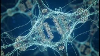
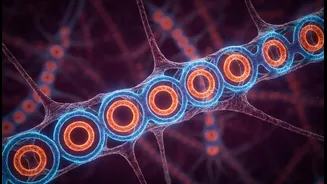
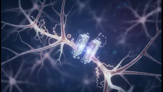
Scientists have identified a lattice-like structure just beneath the surface of neurons called the membrane-associated periodic skeleton, or MPS. For years, it was thought to be a passive scaffold, simply helping the brain cell maintain its shape. However,
new research from Penn State, published in Science Advances, reveals it has a far more active role. Using super-resolution microscopy that can see details 10,000 times smaller than a human hair, researchers found that the MPS acts as a gatekeeper. It actively controls a process called endocytosis, which is how neurons absorb nutrients, proteins, and other materials from their surroundings—a process essential for routine maintenance, learning, and memory. This discovery shifts our understanding from the MPS being a static support to a dynamic regulator of cellular traffic.
The Brain's Remodelling Crew
The MPS is part of the cytoskeleton, a complex network of protein filaments, including actin and microtubules, that provide structure and organization within cells. Think of it as the cell's internal scaffolding and transport system. Ample evidence shows that the cytoskeleton physically reorganises itself in response to learning and memory formation. When we learn, specific connections between neurons, called synapses, are strengthened. This plasticity involves changing the shape of dendritic spines—tiny protrusions on neurons where synapses are located. These physical changes are driven by the actin cytoskeleton. For a memory to last, these new spine structures must be maintained, suggesting the cytoskeleton provides the physical basis for the memory trace itself.
How Stimulation Changes the Structure
Cognitive stimulation—the act of engaging in effortful learning—triggers a cascade of events at the cellular level. This increased neural activity causes the MPS to adapt. The new research found that the MPS can actively break itself down in response to stimulation. When endocytosis, or nutrient uptake, increases, it triggers signals that tell proteins to chop up parts of the skeleton. This creates a feedback loop: increased uptake weakens the lattice, which in turn allows for even more material to enter the cell. This dynamic remodelling allows the neuron to adapt its connections and strengthen the circuits involved in the new learned task. Other studies have described 'actin waves'—growth-cone-like structures that travel down a neuron's projections—which are also linked to neural growth and the competition between developing connections. These waves physically widen the neuron shaft, allowing for increased transport of the materials needed for growth.
Implications for Learning and Brain Health
Understanding how this internal skeleton works has profound implications. On one hand, it provides a physical explanation for how learning and memory are encoded. The idea that our brains physically re-wire themselves isn't just a metaphor; the cell's own skeleton changes. On the other hand, it opens new avenues for understanding neurodegenerative diseases. Conditions like Alzheimer's and Parkinson's are characterized by the harmful buildup of proteins in the brain. The Penn State researchers found that a disrupted MPS allows for the much more rapid absorption of materials, including the amyloid proteins linked to Alzheimer's. This suggests that if the MPS gatekeeper function goes wrong, it could accelerate the disease process. Therefore, finding ways to protect and stabilize this cellular skeleton could offer a new therapeutic strategy to slow the progression of these devastating conditions.