
What's Happening?
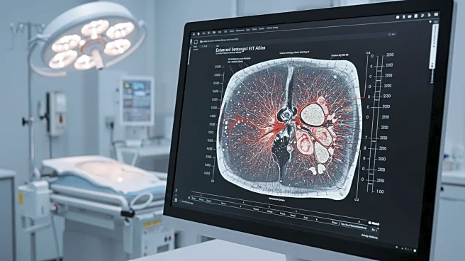
Recent advancements in electrical impedance tomography (EIT) have been made through the development of anatomical atlases constructed from CT scans of infants. These atlases aim to improve real-time reconstructions of ventilation and pulsatile perfusion in preterm infants. The study involved creating two anatomical atlases from CT scans of 89 infants, aged 0 to 3 months, to capture general structures and represent average conductivity and susceptivity tissue values. The atlases were segmented into various tissues using specialized software, and the data was processed to create a comprehensive dataset for EIT imaging. This approach allows for more accurate modeling of infant anatomy, which is crucial for medical applications such as monitoring lung function and diagnosing conditions like bronchopulmonary dysplasia (BPD). The research highlights the potential of using anatomical atlases to enhance the accuracy and efficiency of EIT imaging in clinical settings.
Why It's Important?
The development of anatomical atlases for EIT imaging is significant as it addresses the challenges of accurately reconstructing images of preterm infants' lungs. This advancement could lead to better diagnostic tools for conditions like BPD, which affects many preterm infants. By improving the accuracy of EIT imaging, healthcare providers can monitor lung function more effectively, potentially leading to earlier interventions and improved outcomes for infants with respiratory issues. The use of anatomical atlases also represents a step forward in personalized medicine, as it allows for more tailored imaging based on individual anatomical differences. This could enhance the precision of medical treatments and reduce the risk of complications associated with inaccurate imaging.
What's Next?
The next steps involve further validation of the anatomical atlas approach in clinical settings. Researchers may focus on expanding the dataset to include a broader range of infant anatomies and conditions. Additionally, integrating this technology into existing medical imaging systems could be explored to facilitate its adoption in hospitals and clinics. Collaboration with healthcare providers will be crucial to refine the technology and ensure it meets clinical needs. Future research may also investigate the application of anatomical atlases in other areas of pediatric medicine, potentially broadening its impact beyond respiratory diagnostics.
Beyond the Headlines
The use of anatomical atlases in EIT imaging raises ethical considerations regarding data privacy and consent, especially when dealing with sensitive medical data from infants. Ensuring that data collection and usage comply with ethical standards is essential. Additionally, the technology could influence long-term shifts in pediatric care, emphasizing the importance of personalized and precise medical imaging. As the technology evolves, it may also prompt discussions about the role of machine learning and artificial intelligence in healthcare, particularly in enhancing diagnostic accuracy and efficiency.