


What is the story about?
What's Happening?
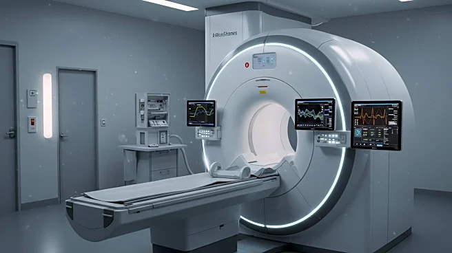
Researchers have developed in-house software to process real-time cine magnetic resonance (MR) images during 1.5 T MR-guided radiation therapy (MRgRT). This software converts binary file formats into metadata or DICOM formats, facilitating image registration and analysis. The study involved 28 imaging sessions across various treatment sites, including prostate and liver, to verify the software's effectiveness. The development aims to enhance the precision of MRgRT by accommodating daily anatomical variations, thereby improving treatment outcomes.
Why It's Important?
The advancement in MR imaging software represents a significant step forward in radiation therapy, offering the potential for more precise and personalized cancer treatments. By improving the accuracy of image registration and analysis, this technology could lead to better targeting of tumors, reducing damage to surrounding healthy tissues. This development is particularly relevant for healthcare providers and patients, as it promises to enhance the effectiveness and safety of radiation therapy.
Beyond the Headlines
The integration of advanced imaging software in radiation therapy could pave the way for broader applications in other medical imaging fields. This innovation may also stimulate further research into real-time imaging technologies, potentially leading to new diagnostic and therapeutic techniques. The ethical and regulatory implications of using such advanced technologies in clinical settings will need to be carefully considered to ensure patient safety and data privacy.