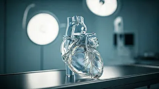
Cardiac Computed Tomography
Cardiac Computed Tomography (CT) scans offer a detailed glimpse into the heart, allowing doctors to assess its overall structure and functionality. This
non-invasive imaging technique creates cross-sectional images of the heart and its surrounding blood vessels. The CT scan is particularly helpful in detecting coronary artery calcium (CAC), a key indicator of atherosclerosis, or the buildup of plaque in the arteries. The scan provides valuable information about the extent of the plaque accumulation, which can help determine the risk of heart disease. The process typically involves lying on a table while the CT scanner rotates around the chest, capturing images. The scan is quick, and the results provide critical insights for preventative care and early intervention strategies to safeguard the heart health.
Electrocardiogram (ECG/EKG)
An Electrocardiogram (ECG or EKG) is a fundamental test used to evaluate the heart's electrical activity. This simple and painless test measures the electrical impulses that control the heartbeat. An ECG can detect various heart problems, including arrhythmias (irregular heartbeats), structural abnormalities, and signs of reduced blood flow to the heart muscle. During an ECG, electrodes are placed on the patient's chest, arms, and legs. These electrodes record the heart's electrical signals, which are then displayed as a series of waves on a monitor or printed on paper. The ECG provides critical diagnostic information, and doctors utilize it to diagnose conditions such as heart attacks, angina, and heart failure. It is a quick and accessible test that offers valuable insights into the heart's function and helps in prompt medical interventions.
Echocardiogram: Sound Waves
An Echocardiogram, often referred to as an echo, utilizes sound waves to create images of the heart, providing detailed information about its structure and function. This non-invasive test allows doctors to assess the size and shape of the heart, the thickness of its walls, and how well it pumps blood. The echocardiogram also examines the heart valves to identify any leakage or narrowing. During the procedure, a technician places a transducer on the patient's chest. This transducer emits ultrasound waves that bounce off the heart, creating real-time images displayed on a monitor. The test aids in the diagnosis of various heart conditions, including valve disorders, cardiomyopathy, and congenital heart defects. An echocardiogram offers valuable insights for diagnosing and managing cardiovascular health, serving as a vital diagnostic tool in cardiology to assess the heart's overall health and function effectively.


