


What's Happening?
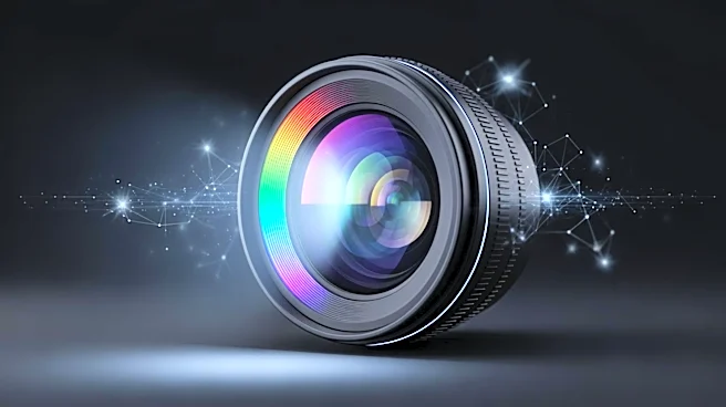
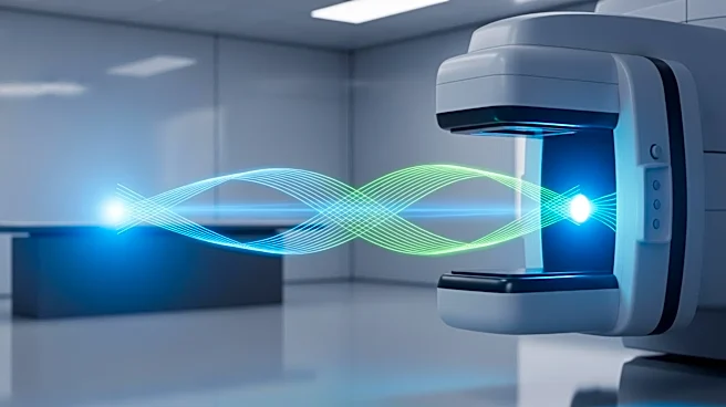
Researchers have developed a non-contact optical imaging model that significantly improves the accuracy of tissue analysis. The model, based on reflectance spectroscopy, eliminates errors caused by direct contact with tissue, enabling precise biochemical
mapping for medical imaging and diagnostics. Published in Scientific Reports, the study introduces a quantitative model for imaging single-fiber reflectance spectroscopy (iSFR), achieving a median prediction error of 6.2%. This advancement allows for reliable extraction of biochemical information without the limitations of traditional fiber-optic probes, which can alter tissue properties and reduce accuracy.
Why It's Important?
The development of a non-contact optical imaging model represents a significant advancement in medical diagnostics. By eliminating the need for direct contact, the model reduces the risk of altering tissue properties, leading to more accurate and reliable results. This technology has the potential to enhance various medical applications, including cancer surgery, where real-time mapping of tissue oxygenation and blood volume can improve surgical outcomes. The integration of this model into existing medical systems could revolutionize how tissue analysis is conducted, offering a safer and more efficient alternative to traditional methods.
What's Next?
Future research will focus on extending the model to more complex tissue structures and a broader range of biological chromophores. The open-source implementation of the model encourages further development by the scientific community, supporting the advancement of non-contact 'optical biopsy' techniques. The integration of this model with other imaging technologies, such as Optical Coherence Tomography (OCT), could provide comprehensive diagnostic tools that combine structural and biochemical analysis. As the model is refined and validated, it may become a standard component in medical imaging, offering new possibilities for non-invasive diagnostics.