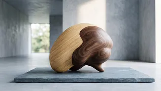








For centuries, we’ve thought of the brain as a single, unified organ. A groundbreaking new study from Stanford Medicine, however, is rewriting the very first chapter of our neural story, revealing our brain may actually be two structures in one.
Our Brain’s Two Main Departments
To understand
this discovery, it helps to know the brain's basic layout. The brain has three main parts, but for this story, we're focused on two. The forebrain is the sophisticated CEO, responsible for uniquely human traits like language, consciousness, and abstract thought. The hindbrain, on the other hand, is the essential operations manager, controlling automatic functions that keep us alive—things like breathing, heartbeat, and sleep. For decades, the prevailing scientific model assumed these vastly different sections all grew from the same single source early in embryonic development.
A Tale of Two Origins
The Stanford research, published in the journal Nature Neuroscience, turns that old model on its head. By studying developing mouse embryos, which provide a powerful model for human development, researchers identified two completely separate populations of 'progenitor' cells. Think of progenitor cells as master cells that are destined to build a specific part of the body. The team found that cells expressing a gene called Otx2 were fated to become the forebrain and midbrain. Meanwhile, a different group of cells expressing a gene called Gbx2 was locked in to forming the hindbrain. Crucially, these two groups of cells never mixed; they developed on parallel tracks that never crossed.
Why This Discovery Is a Game-Changer
This finding does more than just update biology textbooks; it has profound practical implications. For years, scientists have struggled to grow certain types of hindbrain neurons in the lab. This has been a major roadblock in studying devastating neurological diseases that affect the brainstem and hindbrain, such as amyotrophic lateral sclerosis (ALS) and spinal muscular atrophy (SMA). The Stanford study suggests why those attempts failed: researchers were likely trying to coax forebrain-type progenitor cells into becoming hindbrain cells, a transformation the new research shows is impossible. “We've shown for the first time that the front of the brain arises from a totally different progenitor cell than the back of the brain,” said Kyle Loh, an associate professor of developmental biology at Stanford and the study's senior author.
Unlocking New Research Possibilities
Armed with this new knowledge, the researchers were able to successfully grow human hindbrain neurons in a petri dish for the first time. This breakthrough opens up entirely new avenues for research. Scientists can now create accurate lab models to study how hindbrain-specific diseases develop and test potential treatments in ways that were previously out of reach. It’s a fundamental shift from trying to force one cell type to become something it's not, to starting with the correct building blocks from the very beginning. This could accelerate our understanding and ability to combat some of the most challenging neurological conditions.
The Big Question That Remains
While the research answers a long-standing question about the separate origins of the forebrain and hindbrain, it naturally raises another. The headline notes an important question is still open, and while the research itself solves a primary puzzle, the next logical question for the field is a big one: How do these two separately-originating structures, these two 'ancient nervous systems,' integrate so perfectly to form a single, functional brain? The seamless coordination between the part of our brain that thinks and the part that keeps our heart beating is a marvel of biology. Understanding the signals and genetic choreography that stitch these two systems together during development is the next frontier for neuroscientists. The answer will not only deepen our knowledge of evolution but could also provide insights into developmental disorders where this integration may go awry.