




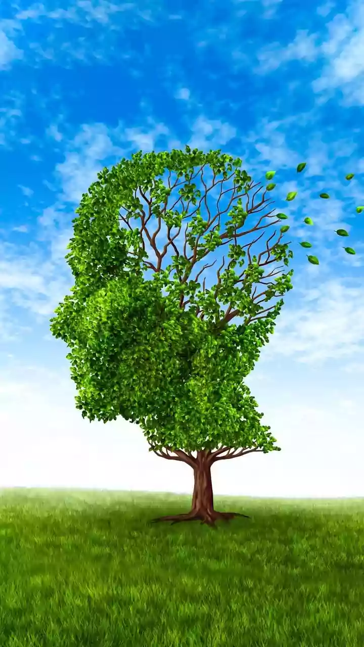



The Old Blueprint for the Brain
For decades, the dominant scientific model of brain development was relatively straightforward. It was widely believed that the entire brain—from the primitive brainstem to the sophisticated cerebral cortex—arose from a single, common group of progenitor
cells in the early embryo. These 'founder' cells were thought to be versatile, capable of differentiating into all the various parts of the brain as development progressed. This model made intuitive sense; after all, the forebrain, midbrain, and hindbrain are all physically connected, appearing to be one contiguous structure. The assumption was that the brain develops much like a tree growing from a single trunk, with different regions branching off as it matures.
A Tale of Two Origins
The new Stanford study, published in Nature Neuroscience, completely overturns this long-held theory. Researchers discovered that the brain is not one system, but two, which are cleverly packaged together. The forebrain, responsible for higher-level functions like language and abstract thought, develops from one set of progenitor cells. The hindbrain, which controls essential automatic functions like breathing, heart rate, and swallowing, arises from an entirely separate and distinct group of cells. These two cell populations are mutually exclusive from the very earliest stages of development, expressing different genes and following parallel tracks that never intersect. It's less like a single tree and more like two different plants whose roots and branches have become intricately intertwined.
How They Discovered It
The breakthrough came from studying mouse embryos at their earliest moments of formation. The scientists identified two types of brain progenitor cells. One type expresses a gene called Otx2, which destines it to become the forebrain and midbrain. The other expresses a gene called Gbx2 and is committed to forming the hindbrain. The team found that the DNA packaging, or chromatin, in these two cell groups was fundamentally different, which effectively locks each cell into its fate. This two-origin pattern was also found in chickens, zebrafish, and even ancient acorn worms, suggesting this developmental blueprint is over 500 million years old.
Why This Is a Game-Changer for Science
This discovery isn't just a fascinating bit of trivia; it solves a decades-long mystery and opens up new fields of research. For years, scientists have struggled to grow certain types of hindbrain neurons in the laboratory. This new finding explains why: they were likely trying to coax forebrain progenitor cells into becoming hindbrain cells, a transformation the research now shows is impossible. One of the study's authors compared it to trying to get to a destination via a concealed side road you never knew existed. Now that researchers understand these two separate starting points, they have successfully grown functional hindbrain neurons from human stem cells for the first time.
New Hope for Neurological Diseases
The ability to grow hindbrain neurons in a lab is a monumental step forward for studying devastating neurological diseases. Many disorders, such as spinal muscular atrophy (SMA) and amyotrophic lateral sclerosis (ALS), specifically affect the hindbrain neurons that control critical functions like swallowing and breathing. Until now, research has been limited by the inability to study these specific human cells. With this new technique, scientists can create models of these diseases in a petri dish, allowing them to investigate how the disorders progress and test potential new therapies without needing tissue from patients. This could dramatically accelerate the search for treatments for some of the most challenging neurological conditions.