






Our Two Brains: The Thinker and The Survivor
To understand this discovery, it helps to know the brain's basic geography. The brain is broadly divided into three parts: the forebrain, midbrain, and hindbrain. The forebrain is the star of the show, responsible for what we consider our 'human' traits:
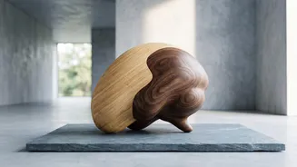
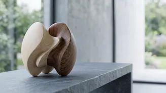
abstract thought, language, and consciousness. The hindbrain, located at the back near the brain stem, is the silent, tireless manager. It handles the essential, automatic functions that keep us alive, such as breathing, heart rate, sleep, and swallowing. For decades, the prevailing model in developmental biology was that all these regions originated from a single, common pool of embryonic cells that later specialized. This new study suggests that model is wrong, proposing instead that the brain is built from two fundamentally different starting blocks.
What the Stanford Study Found
A team led by developmental biologist Kyle Loh at Stanford Medicine investigated the earliest moments of brain development in mouse embryos. They identified two distinct populations of 'progenitor cells' — these are like cellular precursors, destined to become specific types of tissue but not yet fully formed. One group of progenitor cells, which expresses a gene called Otx2, was fated to become the forebrain and midbrain. A second, completely separate group, expressing a gene called Gbx2, was committed to forming the hindbrain. The researchers found that these two cell populations were like travellers on parallel tracks that never cross. They have fundamentally different DNA packaging, known as chromatin, which effectively locks them into their respective developmental paths from the very beginning.
Solving a Decades-Old Frustration
This discovery has immediate practical implications. For years, scientists have struggled to grow certain types of brain cells in the lab, particularly the hindbrain neurons critical for studying devastating diseases. This new research explains why: they were likely using the wrong starter kit. “Previous attempts to make hindbrain neurons likely tried to coax forebrain and midbrain progenitors into hindbrain cells, which our study shows is not possible,” explained Rayyan Jokhai, a co-first author of the study. Armed with this new knowledge, the Stanford team successfully grew functional human hindbrain neurons in a petri dish for the first time. This breakthrough creates a powerful new tool for research.
New Hope for Devastating Diseases
The ability to model the hindbrain in a lab setting is a major step forward for understanding conditions that affect this crucial region. These include spinal muscular atrophy (SMA), a leading genetic cause of death in infants, and amyotrophic lateral sclerosis (ALS), also known as Lou Gehrig's disease. In both disorders, specific hindbrain neurons stop working, leading to a progressive loss of the ability to swallow and, eventually, to breathe. By studying these lab-grown neurons, scientists can now get a much clearer view of how these diseases progress and test potential therapies. The research also has potential connections to understanding obesity, as the hindbrain contains circuits that regulate hunger—the very circuits targeted by popular weight-loss drugs.
An Ancient Evolutionary Story
The finding doesn't just rewrite our understanding of development; it also tells a new story about our evolution. The researchers found the same two-origin pattern in diverse species like chickens, zebrafish, and even acorn worms. This suggests that our brain architecture is ancient, dating back over 500 million years. It appears that evolution took two separate, pre-existing neural systems and elegantly packaged them together to form the complex organ inside our skulls. As Loh puts it, “Having the brain as one organ would probably be more efficient, but we rely on this primordial way to make the brain as two separate pieces.”