
What's Happening?
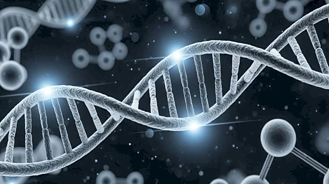
Researchers at Utrecht University have developed a fluorescent sensor that allows scientists to observe DNA damage and repair in real-time within living cells. This breakthrough, detailed in Nature Communications,
provides a continuous view of DNA repair processes, which were previously only observable through isolated snapshots. The sensor uses a fluorescent tag attached to a protein domain that recognizes markers on damaged DNA, enabling researchers to track the entire repair sequence as a continuous movie. This innovation allows for more detailed and realistic observations of cellular processes, potentially revolutionizing cancer biology, drug safety studies, and aging research.
Why It's Important?
The development of this sensor is significant for medical research, particularly in the fields of cancer therapy and drug development. Many cancer treatments rely on inducing DNA damage in tumor cells, and precise measurement of this damage is crucial for evaluating the effectiveness of new drugs. The sensor offers a cheaper, faster, and more accurate method for assessing DNA damage compared to traditional antibodies. Additionally, it can be used to study natural aging processes and detect exposure to mutagenic factors, potentially leading to advancements in understanding and treating various diseases. The tool's availability without restrictions encourages widespread adoption and collaboration among researchers.
What's Next?
The sensor's potential applications extend beyond observing DNA repair. Researchers can use it to map DNA damage locations across the genome and study protein interactions at damaged sites. The tool can also be used in living organisms, as demonstrated in the model organism C. elegans, suggesting its applicability in real-world biological systems. As interest grows, more laboratories are expected to adopt the sensor for their studies, potentially leading to new discoveries in DNA repair mechanisms and their implications for health and disease.