


What's Happening?
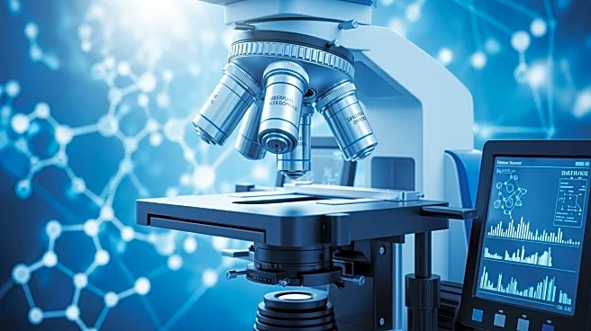
A research team at Westlake University has developed a new method called iPEX, which enables micrometre-resolution deep spatial proteomics through tissue expansion. This innovative approach allows for detailed mapping of protein compositions within tissues,
providing insights into cellular structures and functions. The technique involves expanding tissues using a hydrogel, which facilitates high-resolution imaging and analysis of proteins at a subcellular level. This advancement is expected to significantly enhance the understanding of complex biological systems and disease mechanisms.
Why It's Important?
The development of iPEX is a significant breakthrough in the field of spatial proteomics, offering potential benefits for medical research and diagnostics. By enabling detailed protein mapping, researchers can gain a deeper understanding of cellular processes and disease pathology, which could lead to improved diagnostic tools and targeted therapies. This method may also accelerate research in areas such as cancer, neurodegenerative diseases, and personalized medicine, where understanding protein interactions and distributions is crucial.
What's Next?
The next steps for the research team involve further validation and optimization of the iPEX method for various tissue types and conditions. Collaborations with medical institutions and research centers are likely to expand the application of this technology in clinical settings. Additionally, the team may explore integrating iPEX with other imaging and analytical techniques to enhance its capabilities and broaden its use in biomedical research.
Beyond the Headlines
The iPEX method could have ethical and regulatory implications, particularly in the context of human tissue research. Ensuring compliance with ethical standards and obtaining necessary approvals for clinical applications will be essential. Furthermore, the technology may prompt discussions on data privacy and the management of sensitive biological information.