






What's Happening?
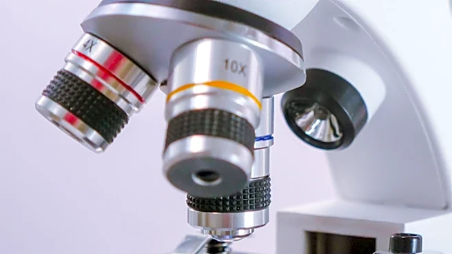
MIT engineers have developed a new microscope capable of imaging electrical activity in neurons across the entire brain of an organism, specifically the zebrafish. This advancement, detailed in Nature Methods, modifies a light sheet microscope to achieve
high-speed, high-volumetric rate imaging. Traditional methods like calcium imaging are too slow to capture individual neuronal impulses, while previous voltage imaging techniques were limited to small populations of neurons. The MIT team enhanced the microscope's image acquisition speed and scanning speed using remote refocusing, allowing it to scan the entire zebrafish brain 200 times per second. This enables researchers to observe millisecond-scale electrical activity, including single voltage spikes and rapid bursts, from neurons distributed throughout the brain. The technique has already revealed patterns of brain activation in response to stimuli like ultraviolet light, showing activity propagating across brain regions such as the optic tectum.
Why It's Important?
This new microscopy technique is crucial for neuroscientists seeking to map neural networks and understand how neural activity underpins behavior and other brain functions. By enabling the observation of electrical impulses across the entire brain at millisecond resolution, it provides an unprecedented view of how different neuron groups coordinate. This holistic approach is vital because brain functions arise from the collective activity of interconnected neurons. The ability to directly observe voltage changes, rather than relying on slower proxies like calcium levels, offers a more accurate and timely understanding of neural computation. This could lead to significant breakthroughs in understanding complex brain processes, from sensory perception and memory formation to movement control, and potentially inform new hypotheses about brain activity during specific behaviors or states of mind like daydreaming.
What's Next?
The MIT researchers plan to further enhance their microscope by increasing the percentage of neurons they can image across the brain, as well as improving the microscope's speed and resolution. They are also working to expand the application of this technique to other experimental models, including mice, which would broaden its utility in neuroscience research. This expansion could provide deeper insights into mammalian brain function and disease. The development of this technology is expected to offer neuroscientists a novel tool to generate and test hypotheses about brain activity, potentially leading to a better understanding of neurological disorders and the development of new therapeutic strategies. Continued refinement of the microscope's capabilities will likely make it an indispensable tool for future brain research.
Beyond the Headlines
The development of this high-speed, whole-brain imaging microscope represents a significant leap in neurotechnology, moving beyond localized studies to a more comprehensive understanding of brain dynamics. This shift towards observing the brain as an 'emergent whole' could fundamentally alter how neuroscientists approach research, emphasizing network-level interactions over individual neuron activity. Ethically, as our ability to map and understand brain activity grows, it raises questions about the implications for understanding consciousness, free will, and the potential for brain-computer interfaces. Legally, advancements in brain imaging could influence areas like mental health diagnostics and forensic neuroscience. Culturally, a deeper understanding of the brain's electrical language could reshape our perceptions of thought, emotion, and behavior, fostering new philosophical and scientific dialogues about the nature of the mind.