
What's Happening?
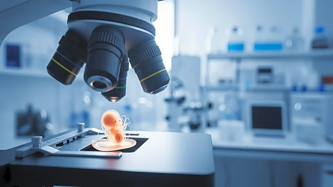
Researchers at the Loke Centre for Trophoblast Research have developed a real-time imaging technique to observe human embryo development, raising questions about the accuracy of current IVF screening methods.
Using light-sheet microscopy, the team observed embryos in 3D, identifying genetic abnormalities that occur late in development. The study found that chromosomal abnormalities often arise in cells destined to become the placenta, which are typically biopsied for preimplantation genetic testing. This discovery suggests that current screening methods may not accurately reflect embryo health, potentially impacting assisted conception practices.
Why It's Important?
The findings from this study could have significant implications for the field of assisted reproduction, particularly in the accuracy of preimplantation genetic testing. The ability to observe embryos in real-time and identify late-stage genetic abnormalities challenges existing screening protocols, which may not account for these errors. This research advocates for a reevaluation of embryo transfer timing and the methods used to assess embryo viability, potentially leading to improved success rates in IVF treatments and better outcomes for families seeking assisted conception.
Beyond the Headlines
The study highlights the importance of direct observation in uncovering unexpected biological phenomena, emphasizing the need for continued research into the causes and consequences of late-stage aneuploidies. The findings may prompt a reconsideration of current clinical practices in IVF, encouraging the development of more accurate and reliable screening techniques. This research also underscores the ethical considerations in assisted reproduction, as it challenges the assumptions underlying embryo selection and genetic testing.