







What's Happening?
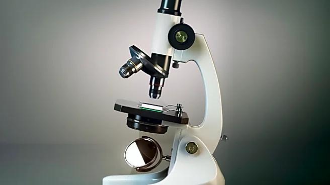
Scientists at the Morgridge Institute for Research have developed an advanced imaging technique, Optical Metabolic Imaging (OMI), capable of characterizing the metabolic activity within individual immune cells from peripheral blood mononuclear cells (PBMCs).
This method, detailed in Biophotonics Discovery, offers a non-destructive way to assess immune cell function, providing more detailed information than traditional cell counts. PBMCs, which are predominantly immune cells, are crucial for understanding disease progression and treatment effects in conditions like certain cancers and immune disorders. The OMI technique utilizes two long-wavelength photons to excite the inherent fluorescence of cell metabolism products, allowing researchers to identify whether PBMCs are metabolically active or quiescent without damaging the cells or requiring chemical labels. This approach achieved 93% accuracy in identifying metabolic states two hours after cell stimulation and could specifically identify quiescent and activated monocytes with 96% and 88% accuracy, respectively, and NK cells with 74% accuracy.
Why It's Important?
This breakthrough in imaging technology holds significant implications for disease diagnostics and the development of cell therapies in the U.S. healthcare system. By providing a more nuanced understanding of immune cell behavior and metabolic states, OMI could lead to improved diagnosis and treatment of immune system-related conditions such as blood cancers, systemic lupus erythematosus (SLE), and sepsis. For CAR T-cell therapy, which engineers a patient's own immune cells to fight cancer, OMI could assess the 'fitness' of these cells before administration, potentially improving patient outcomes. The non-destructive nature of the technique means that samples can be repeatedly imaged over time and then used for downstream applications, preserving valuable patient material. This capability could accelerate research into immune dynamics and facilitate the development of more effective, personalized immune therapies.
What's Next?
The researchers plan to collaborate with hospitals to develop clinical applications for OMI, aiming to integrate this advanced analysis into routine diagnostic and treatment workflows. Efforts are also underway to commercialize the technology, making this label-free metabolic information more accessible to a wider range of laboratories. Future studies will likely focus on validating OMI's utility in larger patient cohorts and across various immune-related diseases. The ability to monitor single-cell behavior over time without destructive manipulation could lead to longitudinal studies that track disease progression and treatment response at an unprecedented level of detail. This could also pave the way for new diagnostic biomarkers based on immune cell metabolic profiles, offering earlier and more precise detection of disease states.
Beyond the Headlines
The development of OMI represents a significant step towards a more personalized and precise approach to medicine, particularly in immunology and oncology. By moving beyond simple cell counts to detailed metabolic profiling, it addresses the inherent heterogeneity of immune responses, which is often masked by bulk measurements. This deeper insight into individual cell behavior could unlock new therapeutic targets and strategies that are currently overlooked. Ethically, the non-destructive nature of OMI is a major advantage, reducing the need for invasive procedures or the use of potentially harmful reagents, thereby improving patient safety and comfort in diagnostic processes. Furthermore, the commercialization efforts highlight the critical bridge between academic research and practical clinical application, emphasizing the potential for advanced imaging techniques to transform healthcare delivery and improve patient outcomes across a spectrum of complex diseases.