






What's Happening?
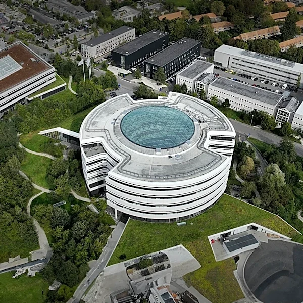
Researchers from the University of Cologne, University Hospital Cologne, and the Max Planck Institute for Metabolism Research have developed a novel computational method to precisely quantify damage to individual cells, moving beyond the traditional approach
of classifying entire organs as healthy or diseased. This technique, detailed in Cell Genomics, utilizes molecular markers, specifically gene expression patterns, to analyze disease progression at a cellular level within tissue samples like biopsies. The method focuses on identifying specific marker genes to measure damage in kidney cells (podocytes) and liver cells (hepatocytes), both crucial in age-related diseases. By applying 'damage scores' to both single-cell RNA sequencing and spatial transcriptomics data, the team can combine molecular identity with information about cellular injury severity and its location within the tissue. This allows for a continuous view of cellular health, ranging from intact to severely damaged, rather than a binary healthy-versus-diseased classification. Professor Dr. Andreas Beyer, who led the study, emphasized the method's universal applicability to other cell types and organs.
Why It's Important?
This new method represents a significant advancement in understanding and treating slowly progressing degenerative diseases, which often develop over years before symptoms become apparent. By providing a high-resolution view of cellular damage, it enables researchers to distinguish early disease mechanisms from later changes and differentiate between patient-specific and general disease progression. This capability is crucial for tailoring treatments more precisely to individual needs, moving towards personalized medicine. The ability to sort cells by damage extent and analyze the sequence of biological processes allows for the identification of critical early stages where interventions would be most effective. This could lead to more targeted therapies and improved patient outcomes in conditions affecting organs like the kidney and liver, which are vulnerable to age-related and degenerative diseases.
What's Next?
The researchers are currently refining the method to enhance its ability to predict disease progression in patients. A more accurate forecast could help identify optimal intervention points and evaluate whether treatments are effectively reversing damage, slowing its spread, or merely managing symptoms. In the long term, these cell-specific damage scores could support the design of clinical studies by enabling investigators to select patients with comparable disease states and measure treatment effects at a molecular level. While not replacing clinical diagnosis, this approach offers a much finer resolution of the processes occurring within diseased tissue. The team plans to adapt the framework to study diseases in other organs, such as the heart, brain, lung, or intestine, by establishing reliable molecular signatures of cellular damage for those specific cell types.
Beyond the Headlines
The development of this computational method signifies a broader shift in biomedical research towards integrating advanced laboratory algorithms with clinical samples. By providing a detailed, cell-by-cell understanding of disease, it challenges the traditional, more generalized view of organ health. This granular insight could revolutionize how degenerative diseases are diagnosed, monitored, and treated, potentially leading to earlier interventions and more effective therapies. The method's compatibility with both single-cell and spatial transcriptomics highlights the growing importance of spatial context in understanding disease, moving beyond simply identifying molecular changes to understanding where and how these changes occur within tissues. This deeper understanding could unlock new pathways for therapeutic development and ultimately improve the quality of life for individuals suffering from chronic degenerative conditions.