


What's Happening?
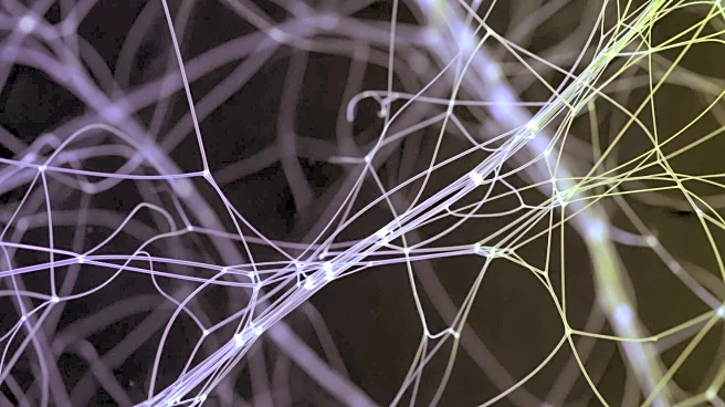
A team of researchers led by Marios Georgiadis, PhD, has developed a new imaging technique called computational scattered light imaging (ComSLI) that uncovers hidden fiber networks within human tissues. This method allows scientists to map the orientation
and organization of tissue fibers at a micrometer resolution on virtually any histology slide, regardless of its age or preservation method. The technique is simple and cost-effective, requiring only a rotating LED light source and a microscope camera. By analyzing how light scatters through microscopic structures, ComSLI generates detailed maps of fiber directions and densities. This breakthrough offers new insights into the microstructure of tissues, which is crucial for understanding health and disease. The research, published in Nature Communications, highlights the potential of ComSLI to transform the study of tissue structure and function across various organs.
Why It's Important?
The development of ComSLI is significant as it addresses the limitations of existing imaging methods, such as MRI and traditional histology, which struggle to capture fine cellular details and resolve complex fiber intersections. By providing a detailed view of tissue microstructures, ComSLI can enhance the understanding of neurological disorders and other diseases where fiber network damage is a factor. This technique opens up new possibilities for research labs and pathology labs to extract valuable data from existing slides, potentially leading to breakthroughs in medical research and diagnostics. The ability to visualize fiber patterns in historical specimens also offers a unique opportunity to study the progression of diseases over time.
What's Next?
The research team plans to expand the application of ComSLI beyond brain research to other tissues such as muscle, bone, and blood vessels. This could lead to a broader understanding of how fiber arrangements contribute to biological functions across different organs. Additionally, there is interest in using ComSLI to revisit well-characterized brain archives and historical specimens to uncover new insights into tissue micro-connectivity. The technique's accessibility and affordability make it a valuable tool for a wide range of scientific and medical communities, potentially leading to new discoveries and advancements in tissue research.
Beyond the Headlines
ComSLI's ability to reveal previously inaccessible data from stored slides worldwide could revolutionize the field of histology. By transforming millions of archived slides into valuable research resources, this technique may lead to a deeper understanding of tissue structure and function. The potential to recover micro-connectivity information from historical specimens, including those of famous individuals, adds an intriguing dimension to the study of human biology and disease. This could provide new perspectives on the evolution of medical conditions and the impact of historical medical practices.