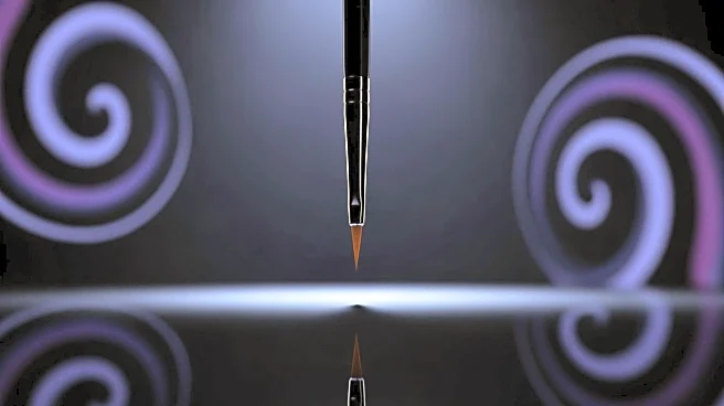


What's Happening?
A research team has made significant progress in the field of single particle imaging by utilizing X-ray free-electron laser (XFEL) technology to retrieve partial orientation of proteins during induced explosions. The study, conducted using Monte Carlo/Molecular
Dynamics simulations, focuses on proteins ranging from 100 to 4000 atoms, which are small enough to allow free electrons to escape during Coulomb explosions. The simulations, carried out with a Gaussian-shaped XFEL pulse, aim to capture the ion trajectories post-explosion using virtual detectors. This method allows researchers to trace ion paths and create a 2D image of the explosion footprint, which is crucial for understanding protein structures. The study highlights the potential of XFEL technology in advancing protein imaging techniques, which could have implications for biological research and drug development.
Why It's Important?
The advancement in protein imaging using XFEL technology is significant for the scientific community, particularly in the fields of biochemistry and molecular biology. Accurate imaging of protein structures is essential for understanding their functions and interactions, which can lead to breakthroughs in drug discovery and development. The ability to retrieve partial orientation of proteins during explosions provides researchers with detailed insights into protein dynamics, potentially accelerating the development of targeted therapies for various diseases. This research also underscores the importance of technological innovation in scientific exploration, paving the way for more precise and efficient methods in studying complex biological systems.