





What's Happening?
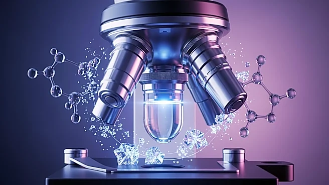
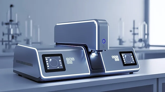
Recent developments in cryogenic electron and mass spectrometry imaging have significantly advanced the field of subcellular chemical mapping. The integration of cryo-EM-FIB-SIMS (cryo-focused ion beam secondary ion mass spectrometry) with existing correlative
light and electron microscopy (CLEM) workflows has enabled researchers to achieve a larger field of view for target identification. This method allows for dual labeling strategies that incorporate both chemical and fluorescent tags, enhancing the ability to visualize and analyze biological specimens at a molecular level. The technique involves the use of gold nanoparticle-tagged bacterial cells and differential fluorescent labeling, which provides detailed spatial and compositional information about subcellular features. The cryo-EM-FIB-SIMS method directly detects secondary ions produced during the interaction between a primary ion beam and the sample, allowing for the detection of elemental, molecular, and isotopic secondary ions. This advancement in imaging technology offers a new dimension of analysis for biological research, particularly in understanding the chemical composition and molecular localization within cells.
Why It's Important?
The integration of cryo-EM-FIB-SIMS into biological research represents a significant leap forward in the ability to map chemical compositions at the subcellular level. This advancement is crucial for various fields, including microbiology, biochemistry, and pharmacology, as it provides a more comprehensive understanding of cellular processes and interactions. The ability to visualize and analyze the chemical makeup of cells with high precision can lead to breakthroughs in drug development, disease diagnosis, and the study of cellular responses to environmental changes. By offering a more detailed view of cellular structures and their chemical environments, this technology can help identify new targets for therapeutic intervention and improve the efficacy of existing treatments. Furthermore, the enhanced imaging capabilities can facilitate the study of complex biological systems, leading to a deeper understanding of life at the molecular level.
What's Next?
As the cryo-EM-FIB-SIMS technology continues to evolve, researchers anticipate further improvements in imaging resolution and compositional sensitivity. Future developments may include the refinement of dual labeling strategies and the expansion of the detectable mass range, which would allow for even more detailed analyses of biological specimens. Additionally, the integration of this technology into routine laboratory workflows could democratize access to high-resolution chemical mapping, enabling a broader range of scientific inquiries. Researchers may also explore the application of this technology in other fields, such as materials science and environmental studies, where understanding the chemical composition at a microscopic level is essential. As the technology becomes more accessible, it is likely to drive innovation across multiple disciplines, leading to new discoveries and applications.
Beyond the Headlines
The advancements in cryo-EM-FIB-SIMS not only enhance scientific research but also raise important ethical and regulatory considerations. As the ability to map chemical compositions at the subcellular level becomes more widespread, questions about data privacy, especially in medical and genetic research, may arise. Ensuring that the technology is used responsibly and ethically will be crucial as it becomes integrated into more areas of research and industry. Additionally, the potential for this technology to uncover previously unknown aspects of cellular biology could lead to paradigm shifts in our understanding of life processes, challenging existing theories and prompting new lines of inquiry. The long-term implications of these discoveries could influence public policy, healthcare practices, and educational curricula, highlighting the need for ongoing dialogue between scientists, ethicists, and policymakers.