


What's Happening?
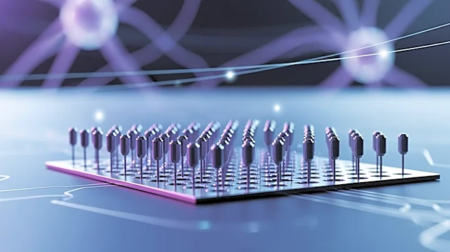
A study using microelectrode arrays (MEAs) has provided new insights into the neuronal and network dynamics in the lateral hypothalamus during sevoflurane anesthesia and emergence. The research involved implanting MEAs in mice to record neuronal activity
across different states of consciousness. The study found distinct firing patterns among neurons, with some showing reduced activity during anesthesia and others exhibiting increased firing. The use of MEAs allowed for detailed observation of neuronal behavior, highlighting the heterogeneous responses of neurons during anesthesia.
Why It's Important?
Understanding neuronal dynamics during anesthesia is crucial for improving anesthetic techniques and patient safety. The findings from this study could lead to better management of anesthesia in clinical settings by providing insights into how different brain regions respond to anesthetic agents. This research also contributes to the broader understanding of brain function and the mechanisms underlying consciousness. The ability to monitor neuronal activity in real-time could enhance the development of new anesthetic drugs and techniques, potentially improving outcomes for patients undergoing surgery.
What's Next?
Further research is needed to explore the implications of these findings for clinical anesthesia. The development of more advanced MEAs and recording techniques could provide even greater insights into brain function during anesthesia. Collaboration between neuroscientists and anesthesiologists will be essential to translate these findings into clinical practice. Additionally, exploring the effects of different anesthetic agents on neuronal dynamics could lead to more personalized and effective anesthesia protocols.