


What's Happening?
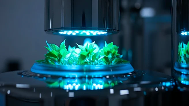
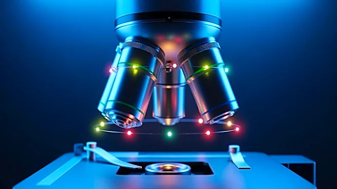
A new technique called brightness demixing has been introduced to improve multi-target imaging in single-molecule localization microscopy. This method allows for the discrimination of fluorophores based on brightness, which is determined by the fluorophore's
extinction coefficient and quantum yield. Unlike traditional methods that rely on spectral separation, brightness demixing uses photon flux to differentiate fluorophores, eliminating the need for additional spectral filters or cameras. This approach enables simultaneous imaging of multiple targets in both two- and three-dimensional configurations, enhancing the capabilities of super-resolution microscopy. The method maintains single-wavelength excitation and minimizes chromatic aberrations, making it compatible with existing microscopy setups.
Why It's Important?
The introduction of brightness demixing in microscopy represents a significant advancement in the field of bioimaging. By allowing for more precise and simultaneous imaging of multiple targets, this method can greatly enhance research in cellular biology and related fields. The ability to differentiate fluorophores without additional spectral separation simplifies the imaging process and reduces equipment costs. This could lead to more widespread adoption of advanced imaging techniques in laboratories, facilitating breakthroughs in understanding complex biological systems. Researchers and institutions stand to benefit from improved imaging capabilities, potentially accelerating discoveries in medical and biological sciences.
What's Next?
As brightness demixing becomes more widely adopted, it is expected that further refinements and applications of this technique will emerge. Researchers may explore its use in various biological contexts, potentially leading to new insights into cellular processes and disease mechanisms. The method's compatibility with existing setups suggests that it could be integrated into current research workflows with minimal disruption. Future developments may focus on enhancing the precision and efficiency of this technique, as well as expanding its applicability to other areas of microscopy and imaging.