





What's Happening?
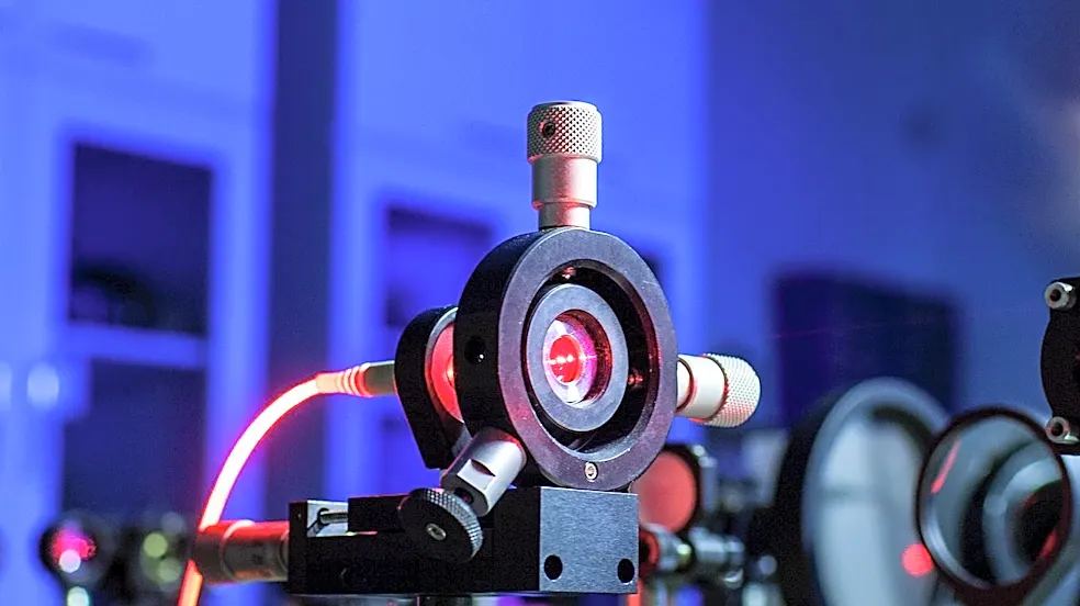
Physicists at the University of California, Berkeley, have developed a groundbreaking device called a laser phase plate, which significantly enhances cryoelectron microscopy (cryo-EM). This innovation, led by UC Berkeley professor of physics Holger Müller,
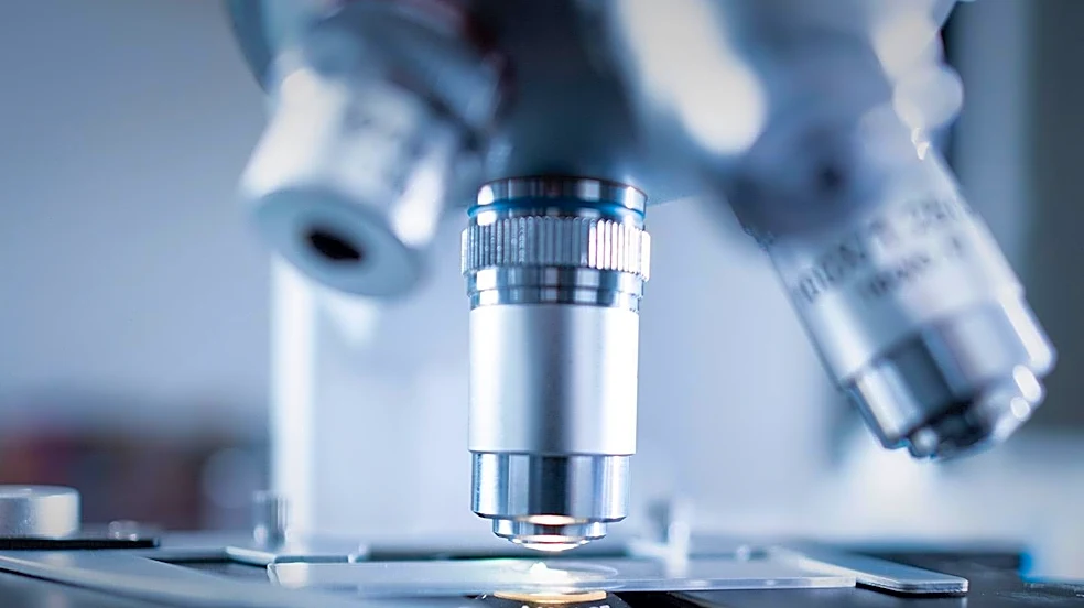
aims to overcome a major limitation in cryo-EM: the difficulty in obtaining clear images of very small proteins. The laser phase plate applies the principle of phase contrast, previously recognized with a Nobel Prize in light microscopy, to electron microscopy, which offers 10,000 times greater magnification. By directly interacting with the microscope's electron beam, the laser alters its phase to increase image contrast, making it easier to distinguish small molecules like hemoglobin and internal cellular structures such as nuclei and mitochondria. This technology is expected to allow scientists to image proteins as small as one-third the size of those currently challenging conventional microscopes. The research, supported by Biohub, has led to the creation of a new microscope named Theia, equipped with this laser phase plate, and a related two-laser system is being developed at Biohub's imaging facility in Redwood City. Both teams are collaborating with Thermo Fisher Scientific, a leading manufacturer of cryo-EM instruments, and their findings are published in the journal Science.
Why It's Important?
This advancement holds profound implications for U.S. biomedical research, drug discovery, and the biotechnology industry. By enabling clearer imaging of smaller proteins, the laser phase plate can unlock a vast, previously inaccessible realm of biological understanding. Approximately 90% of the human proteome consists of proteins smaller than 70 kilodaltons, which conventional cryo-EM struggles to image. This new technology could allow imaging of proteins as small as 50 kilodaltons, potentially expanding to 17 kilodaltons, thereby providing unprecedented insights into the structure and function of a majority of human proteins. This will accelerate drug discovery by allowing researchers to visualize drug targets with greater precision, leading to the development of more effective and targeted therapies. U.S. pharmaceutical companies and biotech firms stand to gain significantly from this enhanced capability, potentially reducing research and development timelines and costs. Furthermore, it could revolutionize our understanding of disease mechanisms at a molecular level, paving the way for novel diagnostic tools and treatments. The collaboration with Thermo Fisher Scientific also suggests a pathway for rapid commercialization and widespread adoption within the scientific community, solidifying the U.S.'s leadership in advanced microscopy and life sciences.
What's Next?
The immediate next steps involve further testing and optimization of the laser phase plate technology, particularly with the two-laser system being developed at Biohub. Researchers will focus on validating its performance across a wider range of biological samples and pushing the limits of resolution for even smaller proteins. Collaboration with Thermo Fisher Scientific will likely intensify to integrate this technology into next-generation commercial cryo-EM instruments, making it accessible to research institutions globally. This could involve developing standardized protocols for its use and training scientists on the new capabilities. Additionally, there will be efforts to explore the full potential of cryoelectron tomography (cryo-ET) with this enhanced contrast, aiming to reconstruct three-dimensional views of proteins within their natural cellular environments. The success of these efforts could lead to increased funding for structural biology research and the establishment of specialized imaging centers equipped with these advanced microscopes. The scientific community will be keenly awaiting further publications detailing the application of this technology to complex biological problems, potentially leading to rapid advancements in various fields of biology and medicine.
Beyond the Headlines
The development of the laser phase plate represents more than just an incremental improvement in microscopy; it signifies a paradigm shift in our ability to visualize the fundamental machinery of life. This technological leap could trigger a cascade of discoveries, fundamentally altering our understanding of cellular processes, disease progression, and the efficacy of therapeutic interventions. Ethically, the enhanced ability to visualize molecular interactions could raise new questions about the definition of life and the potential for manipulating biological systems at an unprecedented scale. Legally, advancements in drug discovery driven by this technology could lead to new intellectual property challenges and regulatory considerations for novel therapies. Culturally, a deeper visual understanding of the molecular world could influence public perception of biology and medicine, fostering greater appreciation for scientific research. In the long term, this technology could democratize access to high-resolution structural biology, enabling a broader range of researchers to tackle complex biological questions, ultimately accelerating the pace of scientific discovery and innovation across the U.S. and globally. It underscores the critical role of fundamental physics research in driving breakthroughs in seemingly disparate fields like biology and medicine.