








What's Happening?
Researchers have developed a 3D-printing method to create miniature models of human blood vessels using CT scans from stroke patients. These models, significantly smaller than actual carotid arteries (reduced to 200-300 micrometers), allow scientists
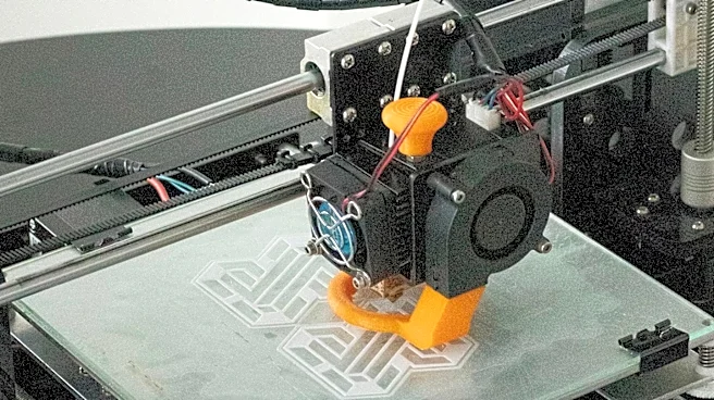
to observe blood flow and platelet behavior under a microscope in real-time. The technology enables the study of how physical forces within irregularly shaped vessels affect platelet movement and clot formation. This research focuses on understanding the processes that contribute to dangerous blood clots, particularly those leading to ischemic strokes. The team found that areas of higher stress within the vessel models led to a 7 to 10 times increase in platelet movement, providing insights into how vessel anatomy and mechanical environment influence clot development. The rapid production time for these models, approximately two hours, facilitates extensive laboratory experimentation.
Why It's Important?
This advancement is crucial for stroke research, offering a novel platform to investigate the complex mechanisms of blood clot formation. By creating patient-specific vascular models, scientists can move beyond generalized studies to understand individual variations in stroke risk and treatment responses. The ability to observe platelet behavior and blood flow dynamics in a controlled laboratory setting addresses a significant challenge in studying clot formation outside the body. This technology could accelerate the development of new diagnostic tools and therapies for stroke, a leading cause of long-term disability and death in the U.S. Understanding how different vessel shapes and blood flow patterns contribute to clotting could lead to more personalized and effective preventative strategies and treatments, ultimately improving patient outcomes and reducing the burden of stroke on the healthcare system.
What's Next?
While currently a research platform, the long-term goal is to combine these physical models with artificial intelligence to develop 'digital twins' of patient blood vessels. This integrated approach could potentially help assess individual stroke risk by analyzing how blood behaves within specific vascular structures. Further research is necessary to determine if laboratory observations from these models reliably correlate with actual events in patients. The researchers aim to expand the application of this technology to investigate how various treatments might affect clot formation. The speed of model creation also suggests the possibility of producing multiple models for comparative studies, which could lead to a deeper understanding of the factors influencing stroke and the efficacy of interventions.
Beyond the Headlines
The development of 3D-printed patient-specific blood vessel models represents a significant step towards personalized medicine in cardiovascular health. This technology not only provides a powerful research tool but also highlights the growing intersection of advanced manufacturing, medical imaging, and biological research. The ethical implications of using patient data to create these 'physical twins' will become increasingly relevant as the technology advances towards clinical applications. Furthermore, the potential for AI integration to predict stroke risk raises questions about data privacy, algorithmic bias, and the responsibility of healthcare providers in communicating such predictions. This innovation could fundamentally change how vascular diseases are studied and treated, shifting from generalized approaches to highly individualized interventions based on a patient's unique anatomy and physiology.