
What's Happening?
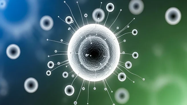
A study by Northwestern Medicine has uncovered the intricate process by which cells coordinate internal scaffolding to form healthy egg cells. Published in the Journal of Cell Biology, the research highlights
the collaboration between actin filaments and microtubules during the development of egg cells in Drosophila melanogaster, commonly known as fruit flies. Actin filaments provide flexibility and support, while microtubules act as rigid tracks for transport and shape. The study reveals that these structures work together to transport cellular supplies to developing egg cells. The research, led by Wen Lu, Ph.D., and Vladimir Gelfand, Ph.D., utilized advanced microscopy techniques to observe the formation of a stable microtubule network and actin cables in nurse cells, which supply nutrients to the egg. The findings demonstrate the essential role of microtubules as a scaffold during oogenesis, the process of egg cell development.
Why It's Important?
Understanding the coordination between actin filaments and microtubules is crucial for comprehending the fundamental processes of egg development, which are conserved across species. This research provides insights into the cellular mechanisms that underpin the development of life, potentially informing future studies in developmental biology and related fields. The study's findings could have implications for understanding fertility and developmental disorders, as well as advancing techniques in reproductive biology. By elucidating the 'crosstalk' between these cellular structures, the research contributes to a deeper understanding of cellular architecture and its role in biological processes.