


What's Happening?
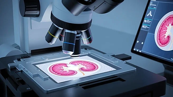
A new label-free optical imaging platform has been developed to improve the analysis of kidney tissue pathology. This platform integrates stimulated Raman scattering (SRS), second harmonic generation (SHG), and multi-photon autofluorescence (MPAF) to capture
detailed spatial morphology and molecular landscapes of kidney tissues. Traditional methods involve serial sectioning and staining, which can consume biopsy volume and risk sample deformation. The new approach offers a nondestructive alternative, providing rich, multimodal views of the same tissue specimen, enhancing diagnostic and research capabilities.
Why It's Important?
The development of this label-free optical imaging platform represents a significant advancement in molecular nephrology. By avoiding the limitations of serial sectioning and staining, this method preserves biopsy integrity and provides more accurate diagnostic information. This can lead to improved treatment decisions and patient outcomes in kidney-related diseases. The integration of multimodal imaging techniques also facilitates the development of deep learning models that can analyze spatial patterns and phenotypes more effectively, potentially revolutionizing pathology research and diagnostics.
Beyond the Headlines
This innovation in optical imaging could have broader implications for other areas of clinical pathology, offering a model for nondestructive analysis of various tissue types. The ability to capture detailed molecular landscapes without damaging samples may lead to new insights in other medical fields, enhancing the overall understanding of disease mechanisms. Additionally, the use of advanced imaging techniques could drive further research into the development of AI models for pathology, improving diagnostic accuracy and efficiency across healthcare.