What's Happening?
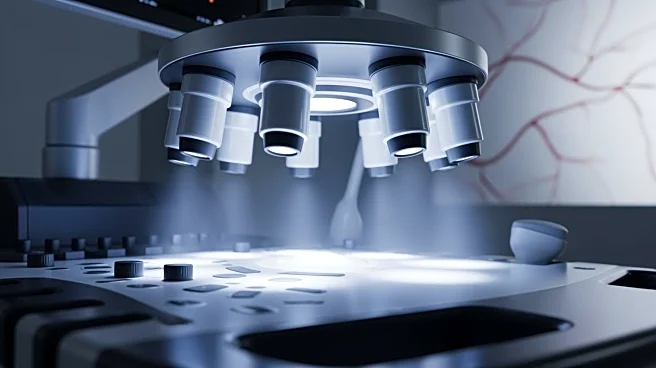
A new ultrasonic multi-lens array approach has been developed to enhance the imaging and quantification of micro-scale vascular networks and flow dynamics in large organs. This method surpasses the limitations
of conventional ultrasound imaging by providing high-resolution volumetric imaging capabilities. Initially validated through simulations and in vitro experiments, the technique was applied to swine organs, demonstrating its ability to visualize entire vascular networks with a spatial resolution down to 125 micrometers. The method also allows for advanced flow dynamic assessments, differentiating arteries and veins and measuring flow velocity. This innovative approach addresses the limitations of existing imaging modalities like ultrasound Doppler and CT angiography by offering a higher spatial resolution and a broader field of view.
Why It's Important?
The development of this multi-lens ultrasound array technique holds significant implications for medical imaging and diagnostics. By providing detailed anatomical and functional insights into vascular structures, it enhances the ability to diagnose and study various conditions, particularly in cardiovascular and chronic diseases. The method's non-invasive nature and real-time capabilities make it a cost-effective alternative to more expensive imaging technologies like MRI. This advancement could lead to improved patient outcomes through more accurate diagnostics and personalized treatment plans. Additionally, the technique's potential applications in preclinical research and drug development could accelerate the understanding and treatment of complex diseases.
What's Next?
The translation of this technology to human applications is anticipated, with potential uses in diagnosing and monitoring chronic kidney and liver diseases. Future developments may focus on optimizing the technique for clinical practice, including improving the microbubble injection protocol and enhancing patient comfort during imaging. The method's adaptability for human use could revolutionize the assessment of organ viability for transplantation and the early detection of diseases. Continued research and development could further refine the technology, making it a standard tool in medical imaging and expanding its applications across various medical fields.
Beyond the Headlines
The introduction of this imaging technique could pave the way for advancements in artificial intelligence and machine learning in medical diagnostics. By generating detailed datasets of organ microcirculation, the method supports the development of digital twins—virtual replicas of patients' physical characteristics—enabling personalized medicine. These digital models could simulate disease progression and treatment outcomes, leading to more effective healthcare solutions. The technique's ability to map complex vascular networks also holds promise for early diagnosis of neurovascular pathologies, potentially transforming the landscape of medical imaging and patient care.


