


What's Happening?
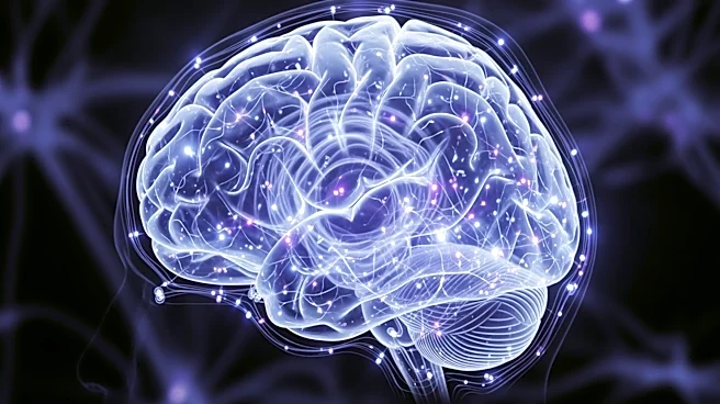
A study published in Communications Biology reveals how the brain processes new faces and places within milliseconds. Using intracranial electroencephalography (iEEG) on patients with epilepsy, researchers tracked brain activity as participants viewed
images of people and places. The study found that different brain regions are activated in a rapid sequence, with faces triggering early responses in occipital and visual stream regions, followed by frontal regions, and later hippocampal and amygdala responses. In contrast, images of places showed earlier hippocampal and amygdala responses. This suggests the brain uses category-specific processing pathways rather than a single visual route, which is crucial for social interactions and memory processing.
Why It's Important?
Understanding how the brain processes visual information is vital for developing treatments for neurological conditions that affect these pathways, such as neurodegenerative disorders and epilepsy. The study's findings could inform future research into perception and the interaction between visual processing, memory, and emotion. By identifying the sequence of brain region activation, scientists can better understand how disruptions in these pathways might occur, potentially leading to improved diagnostic and treatment strategies for conditions that impair visual cognition.
What's Next?
Further research is needed to generalize these findings to larger and more diverse populations. The study highlights the need for advanced imaging techniques that can capture signals at millisecond resolution to better understand the brain's processing of visual information. Future investigations could explore how these pathways are affected in various neurological conditions, potentially leading to new therapeutic approaches.